

The importance of appropriate reporting and investigation of incidental findings on computed tomography attenuation correction images during myocardial perfusion scintigraphy
- PMID: 30774554
- PMCID: PMC6357710
- DOI: 10.4103/wjnm.WJNM_19_18
The importance of appropriate reporting and investigation of incidental findings on computed tomography attenuation correction images during myocardial perfusion scintigraphy
Abstract
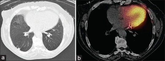
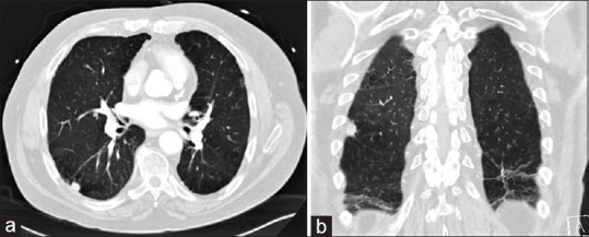
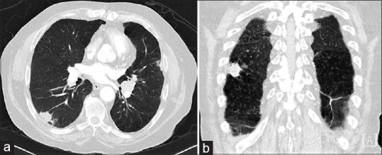
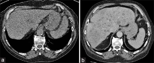
We present a case of lung cancer incidentally detected as a pulmonary nodule on computed tomography attenuation correction (CTAC) images during myocardial perfusion scintigraphy (MPS). Unfortunately, the incidental lesion was not fully investigated following MPS report and had developed into metastatic lung carcinoma when diagnosed over 1 year later, with failure of subsequent emergent chemotherapy. The disease appeared to be localized when initially detected during MPS. This case highlights the importance and potential clinical value of routine review of CTAC images in MPS with appropriate reporting and further investigation of suspicious incidental findings. In addition, the importance of effective communication between nuclear medicine department and treating team is clear to ensure suspicious incidental findings are given sufficient credence and thoroughly investigated promptly to avoid adverse clinical outcomes.
Keywords: Computed tomography attenuation correction; incidental findings; low-dose computed tomography; lung carcinoma; myocardial perfusion scintigraphy.
Conflict of interest statement
There are no conflicts of interest.
Figures
Similar articles
-
Prevalence and clinical significance of incidental findings on CT attenuation correction for myocardial perfusion imaging.J Nucl Cardiol. 2022 Aug;29(4):1813-1822. doi: 10.1007/s12350-020-02499-1. Epub 2021 Mar 22. J Nucl Cardiol. 2022. PMID: 33754302
-
Multi-centre analysis of incidental findings on low-resolution CT attenuation correction images.Br J Radiol. 2014 Oct;87(1042):20130701. doi: 10.1259/bjr.20130701. Epub 2014 Aug 19. Br J Radiol. 2014. PMID: 25135310 Free PMC article.
-
Multicentre analysis of incidental findings on low-resolution CT attenuation correction images: an extended study.Br J Radiol. 2015;88(1056):20150555. doi: 10.1259/bjr.20150555. Epub 2015 Oct 23. Br J Radiol. 2015. PMID: 26493467 Free PMC article.
-
Clinical evaluation of the computed tomography attenuation correction map for myocardial perfusion imaging: the potential for incidental pathology detection.Nucl Med Commun. 2012 Nov;33(11):1122-6. doi: 10.1097/MNM.0b013e3283571b35. Nucl Med Commun. 2012. PMID: 22825039 Review.
-
The Clinical Dilemma of Incidental Findings on the Low-Resolution CT Images from SPECT/CT MPI Studies.J Nucl Med Technol. 2016 Sep;44(3):167-72. doi: 10.2967/jnmt.116.174557. Epub 2016 Apr 21. J Nucl Med Technol. 2016. PMID: 27102662 Review.
Cited by
-
Follow-up of incidental findings on positron emission tomography.World J Nucl Med. 2020 Aug 22;19(3):317-318. doi: 10.4103/wjnm.WJNM_41_20. eCollection 2020 Jul-Sep. World J Nucl Med. 2020. PMID: 33354197 Free PMC article. No abstract available.
References
-
- Pazhenkottil AP, Ghadri JR, Nkoulou RN, Wolfrum M, Buechel RR, Küest SM, et al. Improved outcome prediction by SPECT myocardial perfusion imaging after CT attenuation correction. J Nucl Med. 2011;52:196–200. - PubMed
-
- Goetze S, Pannu HK, Wahl RL. Clinically significant abnormal findings on the “nondiagnostic” CT portion of low-amperage-CT attenuation-corrected myocardial perfusion SPECT/CT studies. J Nucl Med. 2006;47:1312–8. - PubMed
-
- Husmann L, Tatsugami F, Aepli U, Herzog BA, Valenta I, Veit-Haibach P, et al. Prevalence of noncardiac findings on low dose 64-slice computed tomography used for attenuation correction in myocardial perfusion imaging with SPECT. Int J Cardiovasc Imaging. 2009;25:859–65. - PubMed
Publication types
LinkOut - more resources
Full Text Sources