. 2019 Feb 1:9:3181.
doi: 10.3389/fimmu.2018.03181. eCollection 2018.

The Human Endolymphatic Sac and Inner Ear Immunity: Macrophage Interaction and Molecular Expression

Affiliations

The Human Endolymphatic Sac and Inner Ear Immunity: Macrophage Interaction and Molecular Expression

Charlotta Kämpfe Nordström et al. Front Immunol. .

Abstract

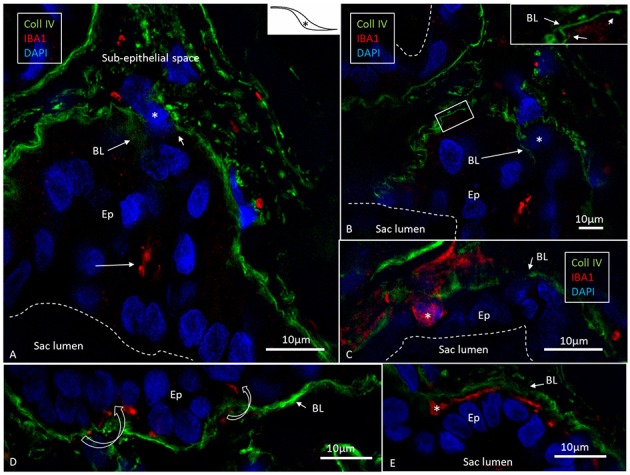
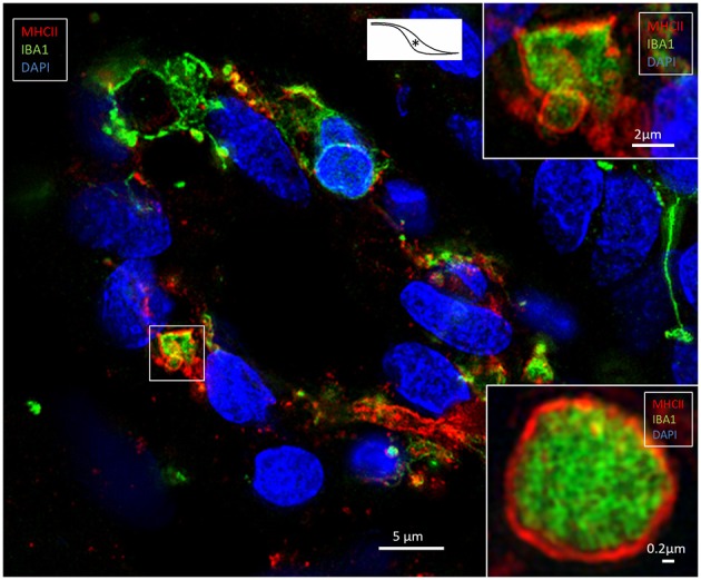
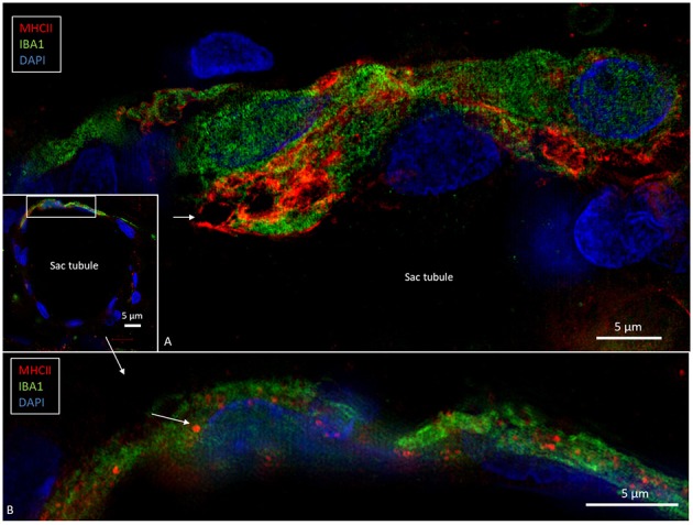
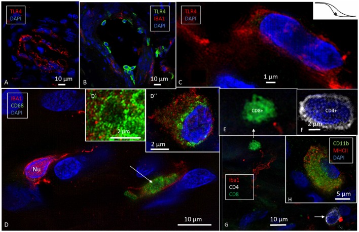
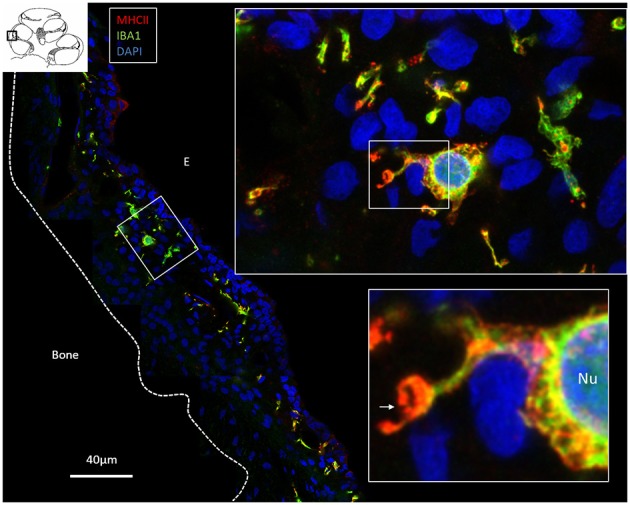
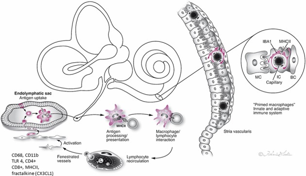
Background: The endolymphatic sac (ES) is endowed with a multitude of white blood cells that may trap and process antigens that reach the inner ear from nearby infection-prone areas, it thus serves as an immunologic defense organ. The human ES, and unexpectedly the rest of the inner ear, has been recently shown to contain numerous resident macrophages. In this paper, we describe ES macrophages using super-resolution structured fluorescence microscopy (SR-SIM) and speculate on these macrophages' roles in human inner ear defense. Material and Methods: After ethical permission was obtained, human vestibular aqueducts were collected during trans-labyrinthine surgery for acoustic neuroma removal. Tissues were placed in fixative before being decalcified, rapidly frozen, and cryostat sectioned. Antibodies against IBA1, cytokine fractalkine (CX3CL1), toll-like receptor 4 (TLR4), cluster of differentiation (CD)68, CD11b, CD4, CD8, and the major histocompatibility complex type II (MHCII) were used for immunohistochemistry. Results: A large number of IBA1-positive cells with different morphologies were found to reside in the ES; the cells populated surrounding connective tissue and the epithelium. Macrophages interacted with other cells, showed migrant behavior, and expressed immune cell markers, all of which suggest their active role in the innate and adaptive inner ear defense and tolerance. Discussion: High-resolution immunohistochemistry shows that antigens reaching the ear may be trapped and processed by an immune cell machinery located in the ES. Thereby inflammatory activity may be evaded near the vulnerable inner ear sensory structures. We speculate on the immune defensive link between the ES and the rest of the inner ear.

Keywords: IBA1; cochlea; human; macrophages; structured illumination microscopy.


Figures

Figure 1
Figure 1
(A) Micro-CT and computer 3D reproduction of a left human inner ear silicon cast. The cochlea (C) and semicircular canals are seen together with the vestibular aqueduct (red). Facial nerve canal is yellow and veins are blue. The vestibular aqueduct (red) houses the endolymphatic duct (ED) and sac. The sac consists of an intraosseous part located inside the temporal bone and an extra-osseous part located on the posterior slope of the petrous pyramid within the dura. (B) The sac exits through an opening in the bone (external aperture of the VA, interrupted line in A,B), and it often extends over the sigmoid sinus (SS). (C) The ED connects the sac with the rest of the inner ear. The intra- and extra-osseous parts of the sac were investigated in the present study. The sac is divided into a proximal (1), an intermediate (2), and a distal part (3). 1 and 3 have a smooth, single-cell layered epithelium, while the intermediate part (2) has a rugose multi-layered epithelium with secretory-like tubules. Demonstration of the bony surface in (B) is possible by using a transparency paradigm within the 3D program. C, Cochlea; JB, Jugular bulb; SS, Sigmoid sinus; IAC, Internal auditory canal; P, Internal acoustic porous; CC, Carotid canal.
Figure 2
Figure 2
SR-SIM (A,B) and confocal fluorescence microscopy (C,D) of the human ES. (A,B) show the difference in nuclear expression of IBA1 protein in macrophages (A) and fibrocytes (B) in the peri-saccular tissue. (C) The sub-epithelial tissue in the intraosseous part of the ES displays a large number of variously shaped IBA1-positive cells. In two of these cells (arrows), the typical nuclear staining can be seen. Several thin cross-sectioned ramifications of IBA1 cells are visible. (D) The epithelium also contains a large number of IBA1 cells, located both between and beneath epithelial cells (inset D′). The epithelial cells express the chemokine fractalkine. *Shows location in the ES.
Figure 3
Figure 3
SR-SIM of human ES near the external aperture of the VA. (A) An elongated sub-epithelial cell expressing IBA1. (B) IBA1-positive cells are located both sub-epithelial and within the epithelium (Ep). One cell seems to have been loosened and expelled into the lumen of the ES. The framed area is magnified in (D). One epithelial cell is surrounded by several IBA1-positive cells (arrows). (C) An IBA1 cell is located next to an epithelial cell (arrow). Sub-epithelial fibrocytes (F) and epithelium express fractalkine. Lv, lymphatics; D, The framed area in B magnified to show an IBA1 cell, presumably being expelled into the lumen; M, Macrophage. *Shows location in the ES.
Figure 4
Figure 4
SR-SIM of epithelial conglomerate at the intra-osseous part of the ES. (A,B) Some epithelial cells (Ep) express the intermediate filament vimentin. (C) The accumulation of IBA1 cells is visible beneath the epithelium and around several vimentin-positive cells. Inset, The IBA1 cells show characteristic nuclear (Nu) staining. *shows location in the ES.
Figure 5
Figure 5
Expression of IBA1 cells and collagen IV. SR-SIM of the intra-osseous part of the ES showing several epithelial tubules. (A) IBA1 cells are located in the epithelium (Ep) as well as in the sub-epithelial space. (B) Adjacent section shows discontinuous basal lamina (BL) and trans-epithelial migration of one IBA1-neagtive cell (*). (C) Large IBA1 cell (*) crosses the BL and epithelium. (D,E) IBA1 cells located between the BL and epithelial cells. Note the extensive fragmentation of BL collagen IV expression in the sub-epithelial space. *Shows location in the ES.
Figure 6
Figure 6
SR-SIM of an epithelial tubule in the intra-osseous part of the ES. Several epithelial cells express IBA1; some also express MHCII. The framed area is magnified in the top inset. The lower inset shows a similar cell, but it is located in the sub-epithelial space. *Shows location in the ES.
Figure 7
Figure 7
SR-SIM of epithelial tubules in the intra-osseous part of the ES. (A) Several IBA1-positive cells are present in the epithelium and seem to constitute an integral part of the epithelium. The cells express MHCII. Large endocytic vesicles (arrow) express MHCII. (B) The framed area of the inset is shown under a higher magnification; some epithelial cells express IBA1 as well as MHCII (arrow).
Figure 8
Figure 8
SR-SIM of cells expressing the toll-like receptor 4 (TLR4, A–C), CD68 (D), and CD11b (H) in the human ES. TLR4 is expressed both in the cell membrane and in the cytoplasm (C). (D) CD68 and IBA1 co-expression in the sub-epithelial cells (arrow, inset). Inset D′ shows CD68+ cell in a higher magnification and D″ shows an IBA1 cell with perinuclear distribution of CD68. (E–G) show sub-epithelial cells expressing lymphocyte markers CD8+ and CD4+ associated with IBA1 cell (arrow, four channels used). (H) One cell co-expresses CD11b and MHCII. *Shows location in the ES.
Figure 9
Figure 9
Confocal microscopy and SIM of the human stria vascularis. Cells co-express IBA1 and MHCII. The framed area is magnified in the insets. Several IBA1 cells are located in the stria epithelium, but few are found in the spiral ligament. Insets, The nuclei (Nu) express IBA1. The cell membrane (arrow) and the cytoplasm contain vesicles expressing MHCII. E, Endolymph.
Figure 10
Figure 10
Hypothetical representation of the immune interaction between the endolymphatic sac (ES) and the inner ear in man. The ES receives antigens and waste material via the endolymphatic duct, which activates resident macrophages/monocytes that then migrate into the sac lumen. After phagocytosis, cells migrate back into the perisaccular tissue for antigen presentation and immune processing. MHC class II molecules, reinforced by TLR4, and under IFNγ stimuli, bind to endocytosed, and degraded peptides within the cytoplasm and transport the peptides to the cell membrane for presentation to CD4+ T cells. The lymphocytes recirculate into the bloodstream to “prime” the inner ear cells. The immune activation of the inner ear is located “off-site” to avoid pro-inflammatory activation near the vulnerable hair cells. The chemokine fractalkine assists in attracting macrophages to the ES (2, 4, 5, 10, 17).

Similar articles

Cited by

References

    1. O'Malley JT, Nadol JB, Jr, McKenna MJ. Anti CD163+, Iba1+, and CD68+ Cells in the adult human inner ear: normal distribution of an unappreciated class of macrophages/microglia and implications for inflammatory otopathology in humans. Otol Neurotol. (2016) 37:99–108. 10.1097/MAO.0000000000000879 - DOI - PMC - PubMed
    1. Liu W, Molnar M, Garnham C, Benav H, Rask-Andersen H. Macrophages in the human cochlea: saviors or predators-a study using super-resolution immunohistochemistry. Front Immunol. (2018) 9:223. 10.3389/fimmu.2018.00223 - DOI - PMC - PubMed
    1. Mei X, Atturo F, Wadin K, Larsson S, Agrawal S, Ladak HM, et al. . Human inner ear blood supply revisited: the Uppsala collection of temporal bone-an international resource of education and collaboration. Ups J Med Sci. (2018) 123:131–42. 10.1080/03009734.2018.1492654 - DOI - PMC - PubMed
    1. Rask-Andersen H, Stahle J. Lymphocyte-macrophage activity in the endolymphatic sac. An ultrastructural study of the rugose endolymphatic sac in the guinea pig. ORL J Otorhinolaryngol Relat Spec. (1979) 41:177–92. 10.1159/000275458 - DOI - PubMed
    1. Rask-Andersen H, Stahle J. Immunodefence of the inner ear? Lymphocyte-macrophage interaction in the endolymphatic sac. Acta Otolaryngol. (1980) 89:283–94. 10.3109/00016488009127140 - DOI - PubMed

Publication types

MeSH terms