

Effects of chitosan scaffold along with royal jelly or bee venom in regeneration of critical sized radial bone defect in rat
- PMID: 30774664
- PMCID: PMC6361593
Effects of chitosan scaffold along with royal jelly or bee venom in regeneration of critical sized radial bone defect in rat
Abstract
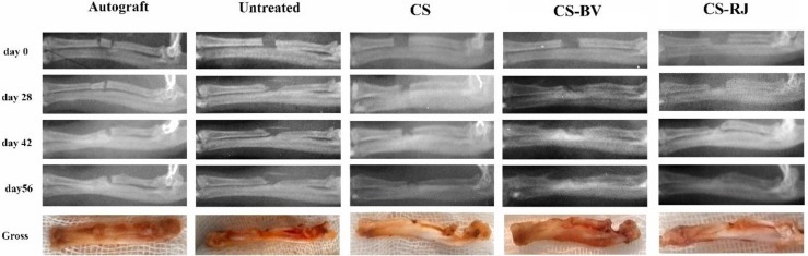
The aim of this study was to compare the efficacy of honey bee venom (BV) and royal jelly (RJ) alongside chitosan scaffold (CS) in improving radius bone defect in rats. A total of 60 full thickness radial bone defects with a length of 5 mm were created in 60 male Wistar rats. Six healthy radial bones (3 rats) were also assigned as normal control for biomechanical studies. The defects were left empty (untreated group) or were filled by the autograft (autograft group), CS (CS group), CS alongside the BV solution (CS-BV group), and CS alongside the RJ solution (CS-RJ group). Healing of the bone defects were evaluated clinically and radiologically on days 0, 28, 42 and 56 after operation while the biomechanical testing and histopathological examination were performed on the 56th day after surgery. The autograft was more radiopaque than the untreated and CS groups at the 28th, 42nd and 56th postoperative days (P<0.05). The CS-BV and CS-RJ groups showed significantly higher radiographic outcomes than the untreated and CS groups at the 56th post-operative day (P<0.05). The density of osseous tissue (DOT) and the osteocytes and osteoblasts count of the CS-RJ and CS-BV groups were significantly higher than the CS and autograft groups (P<0.05). The biomechanical results of the CS-RJ group were significantly superior to the autograft, while the biomechanical properties of CS-BV group were not significantly different with the autograft group (P>0.05). The scaffolds in CS group were observable in the surgical site after 56 days. There was no significant difference in radiographs, DOT, cartilage tissue and fibrous tissue, and also biomechanical performances of the CS-BV and CS-RJ groups at the 42nd and 56th day after surgery. The untreated and CS groups showed weakest biomechanical results among all groups. It could be concluded that both treatment strategies in the CS-BV and CS-RJ groups were appropriate and useful in treating critical bone defects.
Keywords: Bee venom; Bone healing; Chitosan; Royal jelly.
Conflict of interest statement
There is no conflict of interest.
Figures


Similar articles
-
Propolis extract a new reinforcement material in improving bone healing: An in vivo study.Int J Surg. 2018 Aug;56:94-101. doi: 10.1016/j.ijsu.2018.06.006. Epub 2018 Jun 11. Int J Surg. 2018. PMID: 29902525
-
Effectiveness of tissue engineered chitosan-gelatin composite scaffold loaded with human platelet gel in regeneration of critical sized radial bone defect in rat.J Control Release. 2017 May 28;254:65-74. doi: 10.1016/j.jconrel.2017.03.040. Epub 2017 Mar 29. J Control Release. 2017. PMID: 28363521
-
Effectiveness of tissue engineered based platelet gel embedded chitosan scaffold on experimentally induced critical sized segmental bone defect model in rat.Injury. 2017 Jul;48(7):1466-1474. doi: 10.1016/j.injury.2017.04.044. Epub 2017 Apr 24. Injury. 2017. PMID: 28460883
-
The effects of gelatin, fibrin-platelet glue and their combination on healing of the experimental critical bone defect in a rat model: radiological, histological, scanning ultrastructural and biomechanical evaluation.Cell Tissue Bank. 2018 Sep;19(3):341-356. doi: 10.1007/s10561-017-9679-5. Epub 2017 Dec 20. Cell Tissue Bank. 2018. PMID: 29264693
-
Healing potential of nanohydroxyapatite, gelatin, and fibrin-platelet glue combination as tissue engineered scaffolds in radial bone defects of rats.Connect Tissue Res. 2018 Jul;59(4):332-344. doi: 10.1080/03008207.2017.1387541. Epub 2017 Oct 30. Connect Tissue Res. 2018. PMID: 29035127
Cited by
-
Applications of honeybee-derived products in bone tissue engineering.Bone Rep. 2024 Jan 19;20:101740. doi: 10.1016/j.bonr.2024.101740. eCollection 2024 Mar. Bone Rep. 2024. PMID: 38304620 Free PMC article. Review.
-
An overview of de novo bone generation in animal models.J Orthop Res. 2021 Jan;39(1):7-21. doi: 10.1002/jor.24852. Epub 2020 Sep 23. J Orthop Res. 2021. PMID: 32910496 Free PMC article. Review.
-
Pharmacological effects and mechanisms of bee venom and its main components: Recent progress and perspective.Front Pharmacol. 2022 Sep 27;13:1001553. doi: 10.3389/fphar.2022.1001553. eCollection 2022. Front Pharmacol. 2022. PMID: 36238572 Free PMC article. Review.
References
-
- Alidadi S, Oryan A, Bigham-Sadegh A, Moshiri A. Comparative study on the healing potential of chitosan, polymethylmethacrylate, and demineralized bone matrix in radial bone defects of rat. Carbohydr. Polym. 2017;166:236–248. - PubMed
-
- Al Subaie A, Emami E, Tamimi I, Laurenti M, Eimar H, Abdallah MN, Tamimi F. Systemic administration of omeprazole interferes with bone healing and implant osseointegration: an in vivo study on rat tibiae. J. Clin. Periodontol. 2016;43:193–203. - PubMed
-
- Amin MA, Abdel-Raheem IT. Accelerated wound healing and anti-inflammatory effects of physically cross linked polyvinyl alcohol-chitosan hydrogel containing honey bee venom in diabetic rats. Arch. Pharm. Res. 2014;37:1016–1031. - PubMed
-
- Amin M, Abdel-Raheem I, Madkor H. Wound healing and anti-inflammatory activities of bee venom-chitosan blend films. J. Drug Deliv. Sci. Technol. 2008;18:424–430.
-
- Badr G, Hozzein WN, Badr BM, Al Ghamdi A, Saad Eldien HM, Garraud O. Bee venom accelerates wound healing in diabetic mice by suppressing activating transcription factor-3 (ATF-3) and inducible nitric oxide synthase (iNOS)-mediated oxidative stress and recruiting bone marrow-derived endothelial progenitor cells. J. Cell Physiol. 2016;231:2159–2171. - PubMed
LinkOut - more resources
Full Text Sources