

Different cell death types determination in juvenile mice ovarian follicles
- PMID: 30774671
- PMCID: PMC6361600
Different cell death types determination in juvenile mice ovarian follicles
Abstract
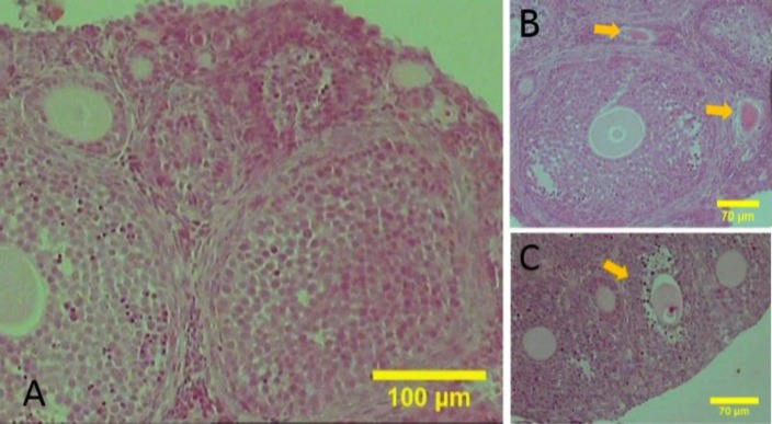
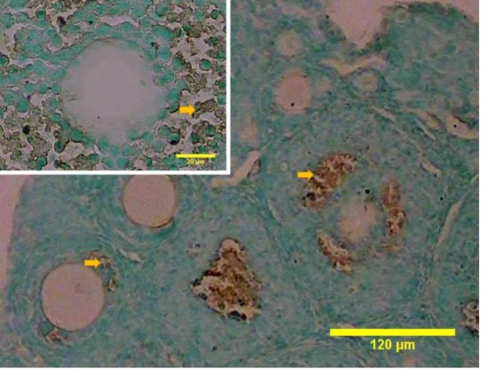
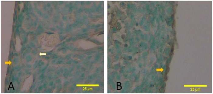
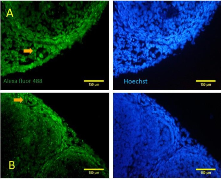
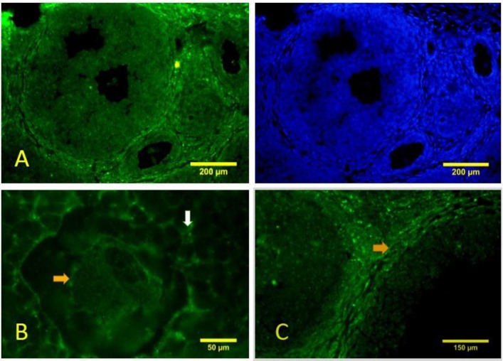
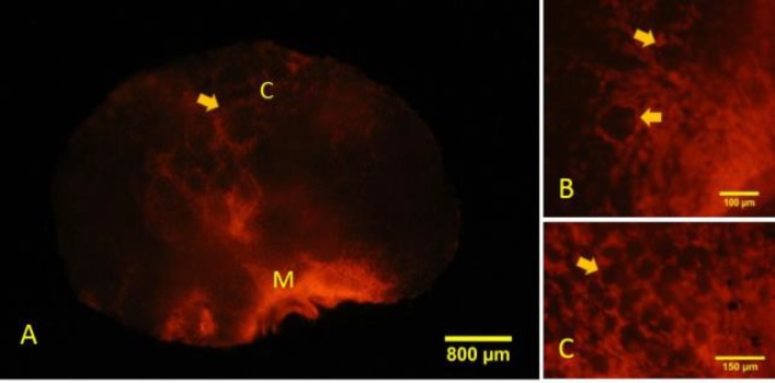
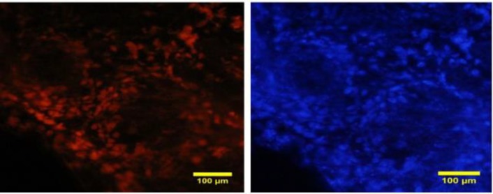
Follicular atresia is a phenomenon that leads to evacuation of the ovary from the oocytes and the occurrence of menopause. The contribution of various types of cell death in atresia at different follicular developmental stages requires extensive investigation. In this study, we evaluated 3 types of programmed cell death (PCD), apoptosis, autophagy, and necrosis, in juvenile mouse ovary when we can observe all follicular stages as well as atresia. Ovaries from juvenile mice on the 21st post-natal (PN) day were prepared histologically for terminal deoxynucleotidyl transferase dUTP nick end labeling (TUNEL) to evaluate apoptosis and immunohistochemistry for beclin-1 to evaluate the autophagy marker. Necrotic cell death was also assessed by penetration of propidium iodide (PI). The count and percentage of the labeled follicles at different stages in the ovaries were evaluated and compared using the Kruskal-Wallis and Mann-Whitney tests. We detected TUNEL-positive granulosa cells in pre-antral and antral follicles but not in the primordial and primary follicles. Somatic cells and oocytes of primordial, primary, pre-antral and antral follicles reacted to beclin-1. The percentage of the PI-labeled primordial and primary follicles were significantly higher than the beclin-1 positive (P=0.01 and P=0.01). In conclusion, we showed that apoptosis, autophagy, and necrosis play a role in follicular atresia and the contributions of each one depends on the follicular stages. It was also demonstrated that necrosis happens particularly in the small follicles while in the large one, all three cell death types occurred with an equal ratio.
Keywords: Apoptosis; Autophagy; Mouse; Necrosis; Ovary.
Conflict of interest statement
The authors declare no conflict of interest.
Figures
Similar articles
-
The infant and pubertal human ovary: Balbiani's body-associated VASA expression, immunohistochemical detection of apoptosis-related BCL2 and BAX proteins, and DNA fragmentation.Hum Reprod. 2013 Mar;28(3):698-706. doi: 10.1093/humrep/des453. Epub 2013 Jan 12. Hum Reprod. 2013. PMID: 23315064
-
Expression of inhibitor of apoptosis proteins (IAPs) in rat granulosa cells during ovarian follicular development and atresia.Endocrinology. 1998 Mar;139(3):1321-8. doi: 10.1210/endo.139.3.5850. Endocrinology. 1998. PMID: 9492068
-
Effects of gonadotropin treatment and withdrawal on follicular growth, cell proliferation, and atresia in ewes.Biol Reprod. 1996 Sep;55(3):693-702. doi: 10.1095/biolreprod55.3.693. Biol Reprod. 1996. PMID: 8862789
-
Role of autophagy in follicular development and maintenance of primordial follicular pool in the ovary.J Cell Physiol. 2022 Feb;237(2):1157-1170. doi: 10.1002/jcp.30613. Epub 2021 Oct 20. J Cell Physiol. 2022. PMID: 34668576 Review.
-
Morphological classification of bovine ovarian follicles.Reproduction. 2010 Feb;139(2):309-18. doi: 10.1530/REP-09-0177. Epub 2009 Sep 28. Reproduction. 2010. PMID: 19786400 Review.
Cited by
-
Folliculogenesis and steroidogenesis alterations after chronic exposure to a human-relevant mixture of environmental toxicants spare the ovarian reserve in the rabbit model.J Ovarian Res. 2024 Jun 28;17(1):134. doi: 10.1186/s13048-024-01457-6. J Ovarian Res. 2024. PMID: 38943138 Free PMC article.
-
A matter of new life and cell death: programmed cell death in the mammalian ovary.J Biomed Sci. 2024 Mar 20;31(1):31. doi: 10.1186/s12929-024-01017-6. J Biomed Sci. 2024. PMID: 38509545 Free PMC article. Review.
References
-
- Amsterdam, A , Sasson, R;Keren-Tal, I , Aharoni, D;Dantes, A;Rimon, E , Land, A , Cohen, T , Dor, Yand Hirsh, L Alternative pathways of ovarian apoptosis: death for life. Biochem. Pharmacol. 2003;66:1355–1362. - PubMed
-
- de Kat AC, Broekmans FJM. Female age and reproductive chances. In: Stoop Dominic., editor. Preventing age related fertility loss. 1st Edn. Switzerland: Springer International Publishing; 2018. pp. 1–10.
-
- D’Herde, K , De Prest, Band Roels, F Subtypes of active cell death in the granulosa of ovarian atretic follicles in the quail (Coturnix coturnix japonica) Rep. Nut. Dev. 1996;36:175–189. - PubMed
-
- Escobar, ML , Echeverría, OM , Ortíz, R , Vázquez-Nin, GH Combined apoptosis and autophagy, the process that eliminates the oocytes of atretic follicles in immature rats. Apoptosis. 2008;13:1253–1266. - PubMed
-
- Gawriluk, TR;Hale, AN , Flaws, JA;Dillon, CP;Green, DR , Rucker, EB Autophagy is a cell survival program for female germ cells in the murine ovary. Reproduction. 2011;141:759–765. - PubMed
LinkOut - more resources
Full Text Sources
Research Materials
Miscellaneous