

Basic and translational neuro-ophthalmology of visually guided saccades: disorders of velocity
- PMID: 30774705
- PMCID: PMC6377082
- DOI: 10.1080/17469899.2017.1395695
Basic and translational neuro-ophthalmology of visually guided saccades: disorders of velocity
Abstract
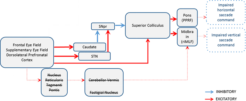
Introduction: Saccades are rapid, yoked eye movements in an effort to direct a target over fovea. The complex circuitry of saccadic eye movements has been exhaustively described. As a result clinicians can elegantly localize the pathology if it falls on the neuraxis responsible for saccades. Traditionally saccades are studied with their quantitative characteristics such as amplitude, velocity, duration, direction, latency and accuracy.
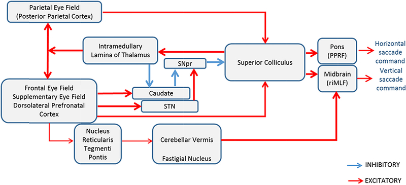
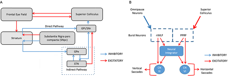
Areas covered: Amongst all subtypes, the physiology of the visually guided saccades is most extensively studied. Here we will review the basic and pertinent neuro-anatomy and physiology of visually guided saccade and then discuss common or classic disorders affecting the velocity of visually guided saccades. We will then discuss the basic mechanism for saccade slowing in these disorders.
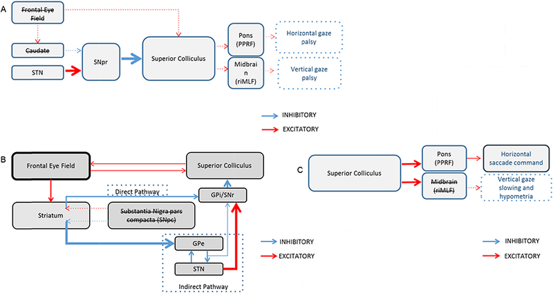
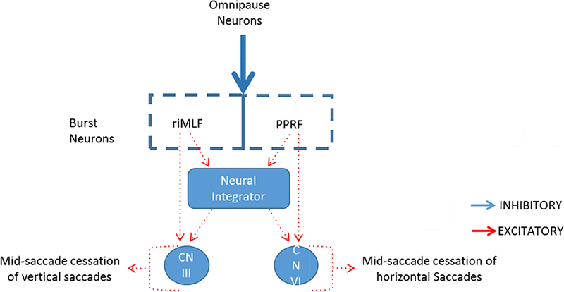
Expert commentary: Prompt appreciation of disorders of saccade velocity is critical to reach appropriate diagnosis. Disorders of midbrain, cerebellum, or basal ganglia can lead to prolonged transition time during gaze shift and decreased saccade velocity.
Keywords: eye movements; saccade; saccade velocity.
Conflict of interest statement
Declaration of interest The authors have no relevant affiliations or financial involvement with any organization or entity with a financial interest in or financial conflict with the subject matter or materials discussed in the manuscript. This includes employment, consultancies, honoraria, stock ownership or options, expert testimony, grants or patents received or pending, or royalties.
Figures
References
-
- Leigh RJ, & Zee DS The neurology of eye movements: Oxford University Press; 2015. Textbook describing basic and applied aspects of eye movements.
-
- DP M Commentary: saccadic eye movements: overview of neural circuitry. Progress in brain research. 2002;140:89–96. - PubMed
-
- Pierrot-Deseilligny C, Milea D, Muri RM. Eye movement control by the cerebral cortex. Curr Opin Neurol. 2004;17:17–25. Comprehensive review on cortical control of eye movements drawing on recent technological advancements in research - PubMed
-
- Fox PT, Fox JM, Raichle ME, Burde RM. The role of cerebral cortex in the generation of voluntary saccades: a positron emission tomographic study. J Neurophysiol. 1985;54:348–69. - PubMed
-
- Pierrot-Deseilligny C, Rivaud S, Gaymard B, Agid Y. Cortical control of reflexive visually-guided saccades. Brain. 1991;114 ( Pt 3):1473–85. - PubMed
Grants and funding
LinkOut - more resources
Full Text Sources