

3D printable hyaluronic acid-based hydrogel for its potential application as a bioink in tissue engineering
- PMID: 30774971
- PMCID: PMC6364434
- DOI: 10.1186/s40824-018-0152-8
3D printable hyaluronic acid-based hydrogel for its potential application as a bioink in tissue engineering
Abstract
Background: After recognition of 3D printing and injectable hydrogel as a critical issue in tissue/organ engineering and regenerative medicine society, many hydrogels as bioinks have been developed worldwide by using polymeric biomaterials such as gelatin, alginate, hyaluronic acid and others. Even though some gels have shown good performances in 3D bioprinting, still their performances do not meet the requirements enough to be used as a bioink in tissue engineering.
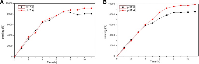
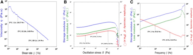
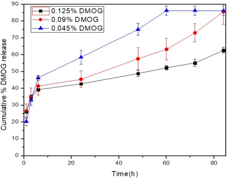
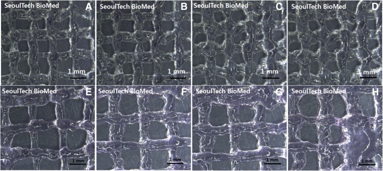
Method: In this study, a hydrogel consisting of three biocompatible biomaterials such as hyaluronic acid (HA), hydroxyethyl acrylate (HEA) and gelatin-methacryloyl, i.e. HA-g-pHEA-gelatin gel, has been evaluated for its possibility as a bioprinting gel, a bioink. Hydrogel synthesis was obtained by graft polymerization of HEA to HA and then grafting of gelatin- methacryloyl via radical polymerization mechanism. Physical and biological properties of the HA-based hydrogels fabricated with different concentrations of methacrylic anhydride (6 and 8%) for gelatin-methacryloylation have been evaluated such as swelling, rheology, morphology, cell compatibility, and delivery of small molecular dimethyloxalylglycine. Printings of HA-g-pHEA-Gelatin gel and its bioink with bone cell loaded in lattice forms were also evaluated by using home-built multi-material (3D bio-) printing system.
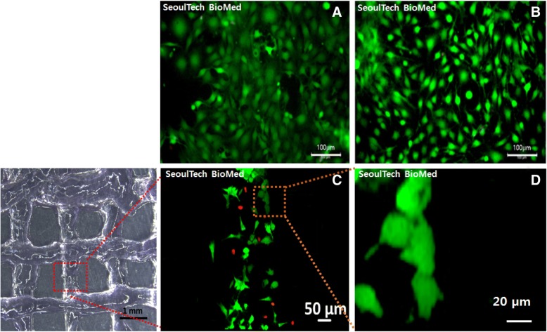
Conclusion: The experimental results demonstrated that the HA-g-pHEA-gelatin hydrogel showed both stable rheology properties and excellent biocompatibility, and the gel showed printability in good shape. The bone cells in bioinks of the lattice-printed scaffolds were viable. This study showed HA-g-pHEA-Gelatin gel's potential as a bioink or its tissue engineering applications in injectable and 3D bioprinting forms.
Keywords: 3D bioprinting; Biocompatible; Gelatin; Hyaluronic acid; Tissue engineering.
Conflict of interest statement
Not applicable.Not applicable.Not applicable.Springer Nature remains neutral with regard to jurisdictional claims in published maps and institutional affiliations.
Figures

References
LinkOut - more resources
Full Text Sources
Other Literature Sources
