. 2019 Feb 8:5:64.
doi: 10.1038/s41420-019-0144-z. eCollection 2019.

The pro-apoptotic Bcl-2 family member Harakiri (HRK) induces cell death in glioblastoma multiforme

Affiliations

The pro-apoptotic Bcl-2 family member Harakiri (HRK) induces cell death in glioblastoma multiforme

Ezgi Kaya-Aksoy et al. Cell Death Discov. .

Abstract

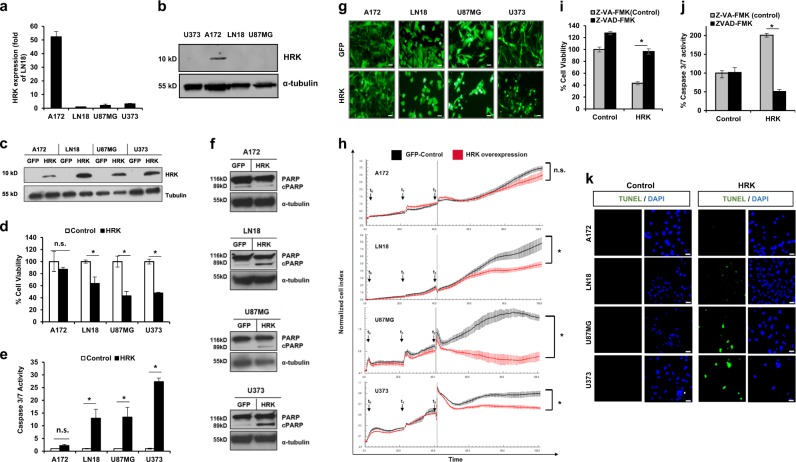
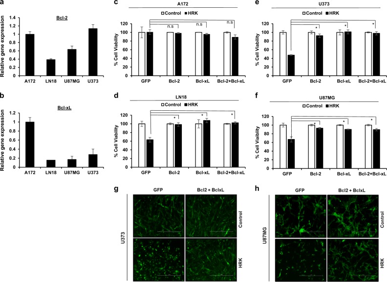
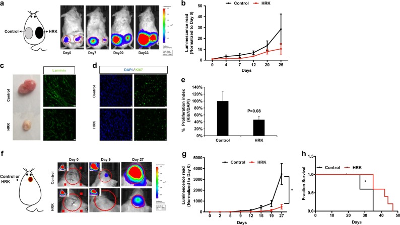
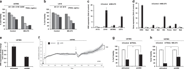
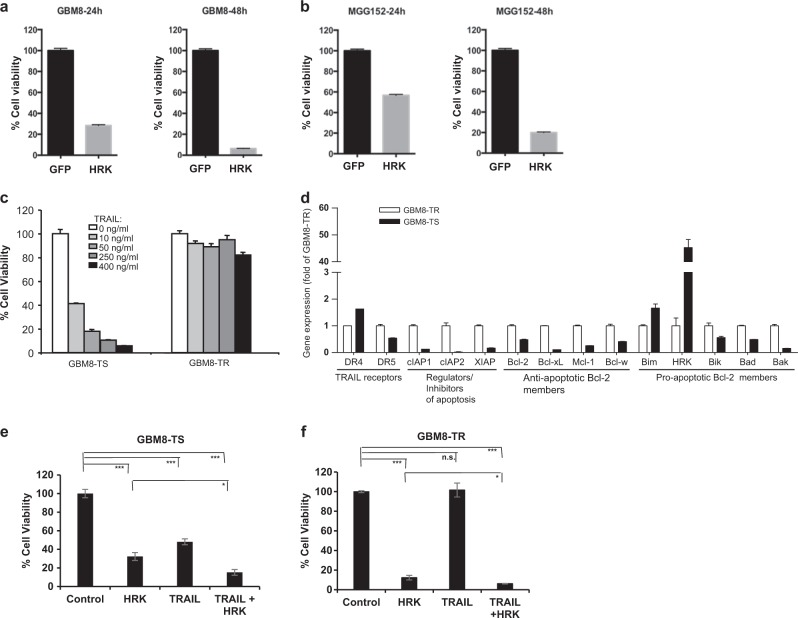
Harakiri (HRK) is a BH3-only protein of the Bcl-2 family and regulates apoptosis by interfering with anti-apoptotic Bcl-2 and Bcl-xL proteins. While its function is mainly characterized in the nervous system, its role in tumors is ill-defined with few studies demonstrating HRK silencing in tumors. In this study, we investigated the role of HRK in the most aggressive primary brain tumor, glioblastoma multiforme (GBM). We showed that HRK is differentially expressed among established GBM cell lines and that HRK overexpression can induce apoptosis in GBM cells at different levels. This phenotype can be blocked by forced expression of Bcl-2 and Bcl-xL, suggesting the functional interaction of Bcl-2/Bcl-xL and HRK in tumor cells. Moreover, HRK overexpression cooperates with tumor necrosis factor-related apoptosis-inducing ligand (TRAIL), a known tumor-specific pro-apoptotic agent. Besides, secondary agents that augment TRAIL response, such as the histone deacetylase inhibitor MS-275, significantly increases HRK expression. In addition, GBM cell response to TRAIL and MS-275 can be partly abolished by HRK silencing. Finally, we showed that HRK induction suppresses tumor growth in orthotopic GBM models in vivo, leading to increased survival. Taken together, our results suggest that HRK expression is associated with GBM cell apoptosis and increasing HRK activity in GBM tumors might offer new therapeutic approaches.


Conflict of interest statement

The authors declare that they have no conflict of interest.

Figures

Fig. 1
Fig. 1. Harakiri overexpression leads to cell death.
a Hrk is differentially expressed in four different established cell lines (A172, LN18, U87MG, U373). Values are normalized to the level of housekeeping gene, GAPDH. b Western blot analysis of endogenous HRK expression in A172, LN18, U87MG, U373 cell lines. c Western blot analysis of HRK in the whole cell lysate of GFP- and HRK-overexpressing GBM cell lines. d, e HRK overexpression decreases cell viability (d) and increases the activation of caspase 3/7 (e). (* denotes p < 0.05, n.s. denotes non-significant (p > 0.05), t-test). f Western blot analysis of PARP cleavage in GFP and HRK-overexpressing GBM cells. g Representative figures showing HRK-induced cell death in GBM cell lines, scale bars:100 µM. h Growth of GFP or HRK-expressing cells in long-term. t0 denotes the time of seeding, t1 denotes the time of viral transduction, t2 denotes the time of medium change. i, j General caspase inhibitor treatment (20 μM ZVAD-FmK or ZVA-FmK) recovers the HRK-induced changes in cell viability (i) and caspase activity (j). k TUNEL staining of GBM cell lines transduced with HRK or control vectors (green: TUNEL, blue: DAPI, scale bars: 10 µM)
Fig. 2
Fig. 2. Bcl-2 and/or Bcl-xL overexpression inhibits HRK-induced death in GBM cells.
a, b Gene expression levels of Bcl-2 and Bcl-xL in A172, LN18, U87MG, and U373 cells detected by qRT-PCR. Values are normalized to the level of housekeeping gene, GAPDH. cf Cell viability effects  of HRK overexpression in GFP, Bcl-2 and/or Bcl-xL overexpressing A172 (c), LN18 (d), U373 (e) and U87MG (f) cells after 48 h HRK transduction. g, h Representative fluorescent images of U373 (g) and U87MG (h) cells transduced with HRK alone (left columns) or together with Bcl-2 and Bcl-xL (right columns) (scale bars:1000 µM) (* denotes p < 0.05, t-test). All experiments were performed in triplicates and representative of technical replicates has been shown
Fig. 3
Fig. 3. HRK overexpression decreases tumor growth in vivo.
a Representative pictures of noninvasive bioluminescence imaging (BLI) of subcutaneous tumors over 33 days. U87MG cells transduced with Luciferase (Fluc)-mCherry (FmC) expressing vectors were post-infected with control or HRK vectors, and then injected subcutaneously into SCID mice (n = 5). b Quantification of tumor growth dynamics by BLI over time. ce) Histological examination of tumors removed at the end of last imaging session. Laminin staining (green) to indicate vascularization (scale bars: 25 µM) (c), Ki67 (green) and DAPI (blue) staining to indicate proliferation and cell nuclei, respectively (scale bars:10 µM) (d), and quantification of proliferation by the number of Ki67-positive puncta over DAPI-positive puncta (n = 5) (e). f) Representative pictures of BLI of intracranial tumor growth over 27 days. U87MG-FmC cells post-transduced with control or HRK vectors were injected intracranially into SCID mice (n = 5 each group) and tumor growth dynamics was measured over time. g) Quantification of tumor growth dynamics by BLI over time. h) Kaplan-Meier survival graphs of intracranial tumor bearing mice implanted with Control- or HRK-expressing cells. (* denotes p < 0.05, ANOVA)
Fig. 4
Fig. 4. HRK overexpression cooperates with TRAIL to induce cell death in GBM cells.
a GBM cells have differential response to TRAIL as measured by cell viability assays of cells in response to TRAIL treatment (0-500 ng/ml) for 24 h. b Cell viability analysis of GFP- or HRK- overexpressing A172, LN18, U87MG, and U373 cells upon 24 h TRAIL treatment (50 ng/ml). c Caspase 3/7 activity analysis of GFP- or HRK- overexpressing A172, LN18, U87MG, U373 GBM cells after 3 h TRAIL treatment (TRAIL concentrations were 20 ng/ml, 20 ng/ml, 50 ng/ml, 200 ng/ml for each cell line respectively). (*, **, *** denote p < 0.05, p < 0.01, p < 0.001, t-test) d, e Effect of HRK and TRAIL cooperation on the growth of U87MG cells in long-term. GBM cells were seeded (t0) and 24 h later were transduced with HRK or GFP viruses (t1). After 24 h, media was refreshed (t2). After 40 h, TRAIL was added (t3) as 25 ng/ml for LN18 (d) and 50 ng/ml TRAIL for U87MG (e) cells (* denotes p < 0.05, ANOVA). fi Western blot analysis of cleaved PARP and Bid protein upon HRK induction and TRAIL treatment (50 ng/ml) in LN18 and U87MG cells. All experiments were performed in triplicates and representative of technical replicates has been shown
Fig. 5
Fig. 5. HRK expression is partly required for TRAIL response and TRAIL sensitization by secondary agents.
a, b Viability analysis of U87MG cells (a) and LN18 cells (b) upon differential doses of TRAIL and MS-275 (5 μM) combination. c qRT-PCR analysis of Hrk expression in four established GBM cell lines (A172, LN18, U87MG, U373) upon MS-275 treatment (5 μM). d qRT-PCR analysis of Bcl-2 family member genes when treated with MS-275 (5 μM). e qRT-PCR analysis of Hrk expression in shControl and shHRK transduced U87MG cells. Values are normalized to the level of housekeeping gene, GAPDH. f Long-term cell growth analysis of shControl and shHRK U87MG cells (t0: time of cell seeding, t1: time of media change). g Cell viability analysis of shControl and shHRK transduced U87MG cells upon TRAIL treatment (75 ng/ml). h Cell viability analysis of shControl and shHRK transduced U87MG cells upon TRAIL (75 ng/ml) and MS-275 (5 μM) combination. (*, **, *** denote p < 0.05, p < 0.01, p < 0.001, t-test). All experiments were performed in triplicates and representative of technical replicates has been shown
Fig. 6
Fig. 6. HRK is differentially expressed in different GBM cell subpopulations and cooperates with TRAIL in primary GBM cell lines.
a, b HRK overexpression decreases the viability of primary GBM cell lines, GBM8 (a) and MGG152 (b), in a time-dependent manner. c TRAIL response of an isogenic GBM cell line (GBM8) pair selected for TS (TRAIL sensitive) and TR (TRAIL resistance) subpopulations that were exposed with differential doses of TRAIL (0, 10, 50, 250, 400 ng/mL) were assessed. d qRT-PCR analysis of apoptotic members in GBM8-TS and GBM8-TR subpopulations was performed. e, f HRK overexpression decreases cell viability and cooperates with TRAIL in TRAIL-resistant (e) and TRAIL-sensitive (f) subpopulation of a primary GBM cells. (n.s., *, *** denote non-significant, p < 0.05, p < 0.001, respectively, t-test)

Similar articles

Cited by

References

    1. Sathornsumetee S, et al. Molecularly targeted therapy for malignant glioma. Cancer. 2007;110:13–24. doi: 10.1002/cncr.22741. - DOI - PubMed
    1. Bonavia R, Inda MDM, Cavenee WK, Furnari FB. Heterogeneity maintenance in glioblastoma: a social network. Cancer Res. 2011;71:4055–4060. doi: 10.1158/0008-5472.CAN-11-0153. - DOI - PMC - PubMed
    1. Furnari FB, et al. Malignant astrocytic glioma: genetics, biology, and paths to treatment. Genes Dev. 2007;21:2683–2710. doi: 10.1101/gad.1596707. - DOI - PubMed
    1. Falschlehner C, Emmerich CH, Gerlach B, Walczak H. TRAIL signalling: decisions between life and death. Int. J. Biochem. Cell Biol. 2007;39:1462–1475. doi: 10.1016/j.biocel.2007.02.007. - DOI - PubMed
    1. Lemke J, von Karstedt S, Zinngrebe J, Walczak H. Getting TRAIL back on track for cancer therapy. Cell Death Differ. 2014;21:1350–1364. doi: 10.1038/cdd.2014.81. - DOI - PMC - PubMed