

Three cell deaths and a funeral: macrophage clearance of cells undergoing distinct modes of cell death
- PMID: 30774993
- PMCID: PMC6368547
- DOI: 10.1038/s41420-019-0146-x
Three cell deaths and a funeral: macrophage clearance of cells undergoing distinct modes of cell death
Abstract
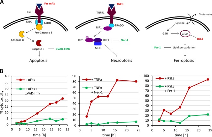
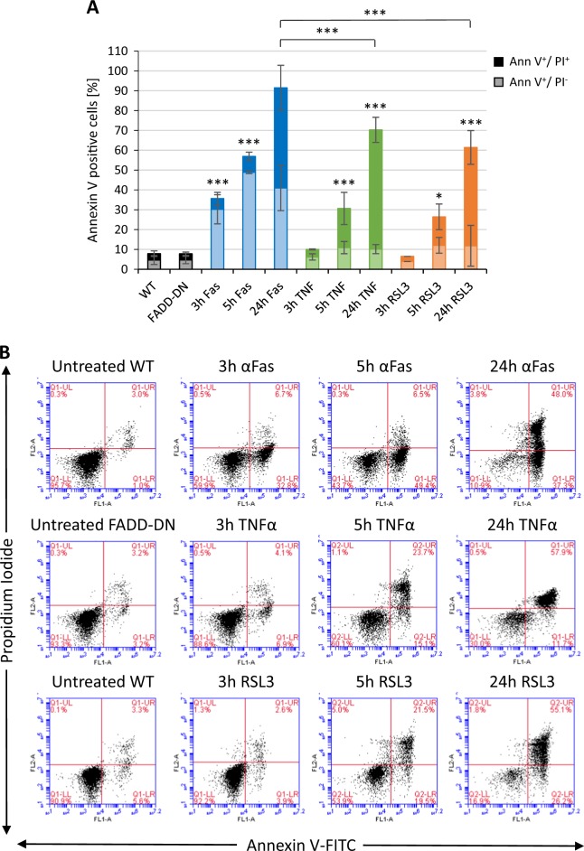
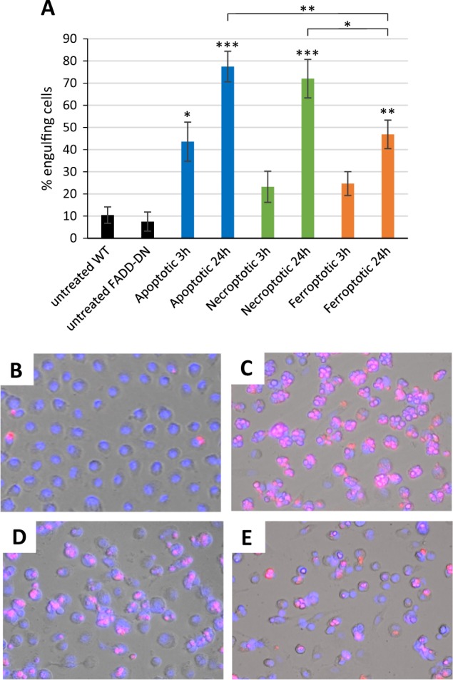
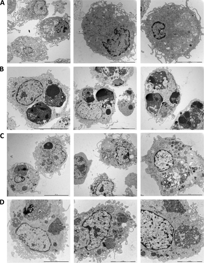
Macrophage clearance of apoptotic cells has been extensively investigated, but less is known regarding the clearance of cells dying by other forms of programmed cell death, e.g., necroptosis or ferroptosis. Here, we established a model of three different cell deaths using the same cell line and the occurrence of distinct cell death modalities was verified by using the specific inhibitors, zVAD-fmk, necrostatin-1, and ferrostatin-1, respectively. Cell death was characterized by using transmission electron microscopy (TEM), the gold standard for the demarcation of different cell death modalities. Moreover, using annexin V as a probe, we could detect surface exposure of phosphatidylserine (PS) in all three types of cell death, and this was confirmed by using specific anti-PS antibodies. We then co-cultured the cells with human monocyte-derived macrophages and found that cells dying by all three death modalities were engulfed by macrophages. Macrophage clearance of apoptotic cells was more efficient when compared to necroptotic and ferroptotic cells with multiple internalized target cells per macrophage, as shown by TEM. We propose that clearance of dying cells also should be taken into account in the classification of different cell death modalities.
Conflict of interest statement
The authors declare that they have no conflict of interest.
Figures
Similar articles
-
Immunogenic cell death induced by a new photodynamic therapy based on photosens and photodithazine.J Immunother Cancer. 2019 Dec 16;7(1):350. doi: 10.1186/s40425-019-0826-3. J Immunother Cancer. 2019. PMID: 31842994 Free PMC article.
-
Pan-caspase inhibitors induce necroptosis via ROS-mediated activation of mixed lineage kinase domain-like protein and p38 in classically activated macrophages.Exp Cell Res. 2019 Jul 15;380(2):171-179. doi: 10.1016/j.yexcr.2019.04.027. Epub 2019 Apr 27. Exp Cell Res. 2019. PMID: 31039349
-
Lactadherin detects early phosphatidylserine exposure on immortalized leukemia cells undergoing programmed cell death.Cytometry A. 2006 Dec 1;69(12):1193-201. doi: 10.1002/cyto.a.20345. Cytometry A. 2006. PMID: 17123296
-
Programmed necrotic cell death of macrophages: Focus on pyroptosis, necroptosis, and parthanatos.Redox Biol. 2019 Sep;26:101239. doi: 10.1016/j.redox.2019.101239. Epub 2019 Jun 9. Redox Biol. 2019. PMID: 31212216 Free PMC article. Review.
-
Friendly fire against neutrophils: proteolytic enzymes confuse the recognition of apoptotic cells by macrophages.Biochimie. 2008 Feb;90(2):405-15. doi: 10.1016/j.biochi.2007.09.008. Epub 2007 Sep 22. Biochimie. 2008. PMID: 17964056 Review.
Cited by
-
Ferroptosis in tumor immunity and therapy.J Cell Mol Med. 2022 Nov;26(22):5565-5579. doi: 10.1111/jcmm.17529. Epub 2022 Nov 1. J Cell Mol Med. 2022. PMID: 36317423 Free PMC article. Review.
-
COVID-19 virtual patient cohort suggests immune mechanisms driving disease outcomes.PLoS Pathog. 2021 Jul 14;17(7):e1009753. doi: 10.1371/journal.ppat.1009753. eCollection 2021 Jul. PLoS Pathog. 2021. PMID: 34260666 Free PMC article.
-
COVID-19 virtual patient cohort reveals immune mechanisms driving disease outcomes.bioRxiv [Preprint]. 2021 Jan 6:2021.01.05.425420. doi: 10.1101/2021.01.05.425420. bioRxiv. 2021. Update in: PLoS Pathog. 2021 Jul 14;17(7):e1009753. doi: 10.1371/journal.ppat.1009753. PMID: 33442689 Free PMC article. Updated. Preprint.
-
Severe congenital neutropenia-associated JAGN1 mutations unleash a calpain-dependent cell death programme in myeloid cells.Br J Haematol. 2021 Jan;192(1):200-211. doi: 10.1111/bjh.17137. Epub 2020 Nov 18. Br J Haematol. 2021. PMID: 33206996 Free PMC article.
-
Non-apoptotic regulated cell death mediates reprogramming of the tumour immune microenvironment by macrophages.J Cell Mol Med. 2024 Apr;28(8):e18348. doi: 10.1111/jcmm.18348. J Cell Mol Med. 2024. PMID: 38652105 Free PMC article. Review.
References
LinkOut - more resources
Full Text Sources
