. 2018 Dec 7;10(1):104-118.
doi: 10.1364/BOE.10.000104. eCollection 2019 Jan 1.

Biophysical basis of skin cancer margin assessment using Raman spectroscopy

Affiliations

Biophysical basis of skin cancer margin assessment using Raman spectroscopy

Xu Feng et al. Biomed Opt Express. .

Abstract

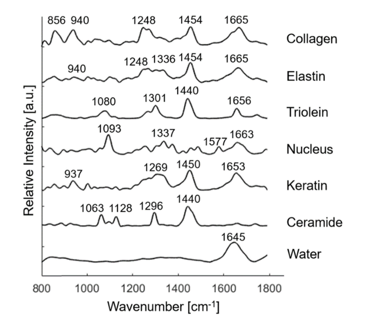
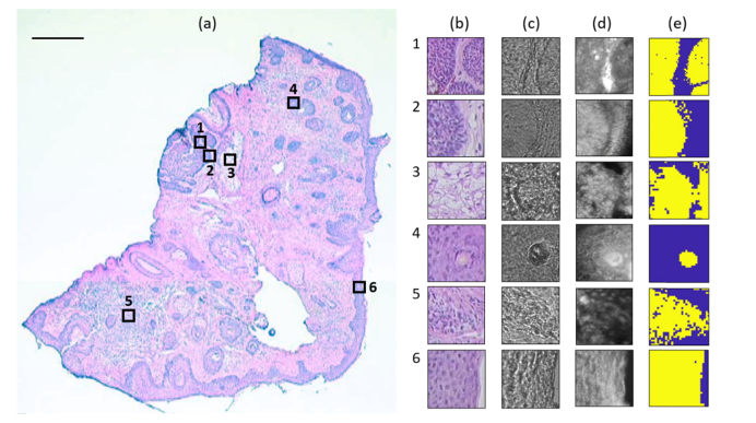
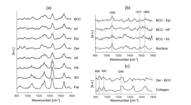
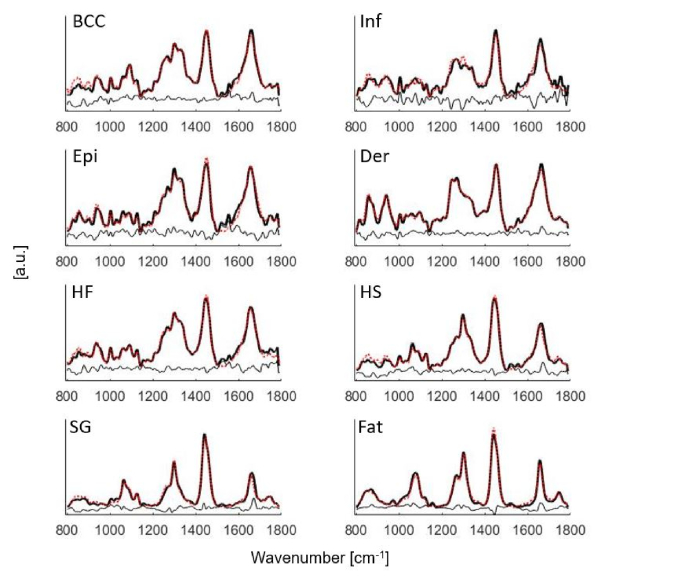
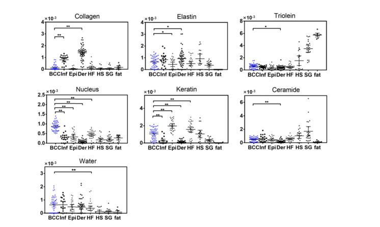
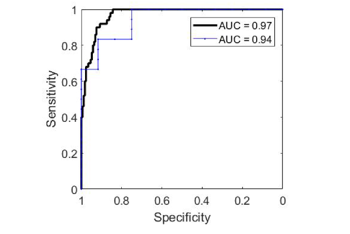
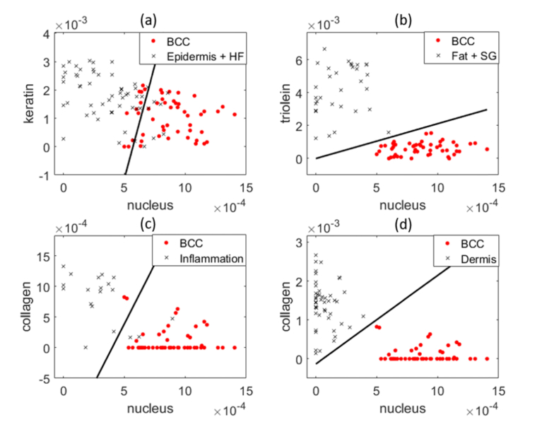
Achieving adequate margins during tumor margin resection is critical to minimize the recurrence rate and maximize positive patient outcomes during skin cancer surgery. Although Mohs micrographic surgery is by far the most effective method to treat nonmelanoma skin cancer, it can be limited by its inherent required infrastructure, including time-consuming and expensive on-site histopathology. Previous studies have demonstrated that Raman spectroscopy can accurately detect basal cell carcinoma (BCC) from surrounding normal tissue; however, the biophysical basis of the detection remained unclear. Therefore, we aim to explore the relevant Raman biomarkers to guide BCC margin resection. Raman imaging was performed on skin tissue samples from 30 patients undergoing Mohs surgery. High correlations were found between the histopathology and Raman images for BCC and primary normal structures (including epidermis, dermis, inflamed dermis, hair follicle, hair shaft, sebaceous gland and fat). A previously developed model was used to extract the biochemical changes associated with malignancy. Our results showed that BCC had a significantly different concentration of nucleus, keratin, collagen, triolein and ceramide compared to normal structures. The nucleus accounted for most of the discriminant power (90% sensitivity, 92% specificity - balanced approach). Our findings suggest that Raman spectroscopy is a promising surgical guidance tool for identifying tumors in the resection margins.


Conflict of interest statement

The authors declare that there are no conflicts of interest related to this article.

Figures

Fig. 1
Fig. 1
Raman active components in the biophysical model of skin, including collagen, elastin, triolein, nucleus, keratin, ceramide, and water. Peak positions of the main Raman bands are labeled.
Fig. 2
Fig. 2
Raman experiment on a typical skin tissue section. (a) H&E image shows six measured regions of 100 × 100 μm2, being represented as empty squares. Scale bar: 500 μm. (b) H&E image of the serial section. (c) Bright-field image. (d) Reflectance confocal images. (e) Raman pseudo-color image generated by k-means. Region 1 and 2 contains BCC (yellow) and dermis (blue), region 3 contains sebaceous gland (yellow) and MgF2 substrate (blue), region 4 contains hair shaft (yellow) and hair follicle (blue), region 5 contains inflamed dermis (yellow) and dermis (blue), and region 6 contains epidermis (yellow) and MgF2 substrate (blue).
Fig. 3
Fig. 3
(a) Mean Raman spectra ± SD of all individual tissue structures, including BCC, Inf (inflamed dermis), Epi (epidermis), Der (dermis), HF (hair follicle), HS (hair shaft), SG (sebaceous gland) and fat. (b) Spectral differences of mean spectra of BCC minus Epi, BCC minus HF, and BCC minus Inf are compared with the basis spectrum of nucleus. (c) Spectral difference of mean spectra of dermis minus BCC is compared with the basis spectrum of collagen. Peak positions of the main Raman bands are labeled.
Fig. 4
Fig. 4
Mean Raman spectra of BCC, Inf (inflamed dermis), Epi (epidermis), Der (dermis), HF (hair follicle), HS (hair shaft), SG (sebaceous gland) and fat fit to the model components in Fig. 1. Black solid lines: mean tissue spectra. Red dotted lines: model fits. Residuals are also plotted on the bottom.
Fig. 5
Fig. 5
Fit coefficients of the biophysical markers for BCC (N = 50), Inf (inflamed dermis, N = 19), Epi (epidermis, N = 26), Der (dermis, N = 47), HF (hair follicle, N = 31), HS (hair shaft, N = 18), SG (sebaceous gland, N = 22) and fat (N = 10). Each point represents a spectrum data. Statistical significance of BCC versus Inf, BCC versus Epi, BCC versus Der, and BCC versus HF are labeled. *p≤0.05, **p≤0.01.
Fig. 6
Fig. 6
ROC analysis for classifying BCC from all normal structures. Black thick line: leave-one-spectrum-out ROC curve. Blue thin line: leave-one-patient-out ROC curve.
Fig. 7
Fig. 7
Scatter plots demonstrates the performance of two primary model components in discriminating BCC from normal structures. (a) Nucleus and keratin content of BCC, epidermis and HF. (b) Nucleus and triolein content of BCC, fat and SG. (c) Nucleus and ceramide content of BCC and inflamed dermis. (d) Nucleus and collagen content of BCC and dermis. Red dots: BCC. Black crosses: normal tissue structures. Each point represents a spectrum data. The black line is the decision line drawn by logistic regression.

References

    1. Society A. C., Cancer facts & figures 2018 (American Cancer Society, Altanta, 2018).
    1. Mosterd K., Krekels G. A., Nieman F. H., Ostertag J. U., Essers B. A., Dirksen C. D., Steijlen P. M., Vermeulen A., Neumann H., Kelleners-Smeets N. W., “Surgical excision versus Mohs’ micrographic surgery for primary and recurrent basal-cell carcinoma of the face: a prospective randomised controlled trial with 5-years’ follow-up,” Lancet Oncol. 9(12), 1149–1156 (2008).10.1016/S1470-2045(08)70260-2 - DOI - PubMed
    1. Smeets N. W., Kuijpers D. I., Nelemans P., Ostertag J. U., Verhaegh M. E., Krekels G. A., Neumann H. A., “Mohs’ micrographic surgery for treatment of basal cell carcinoma of the face--results of a retrospective study and review of the literature,” Br. J. Dermatol. 151(1), 141–147 (2004).10.1111/j.1365-2133.2004.06047.x - DOI - PubMed
    1. Viola K. V., Jhaveri M. B., Soulos P. R., Turner R. B., Tolpinrud W. L., Doshi D., Gross C. P., “Mohs micrographic surgery and surgical excision for nonmelanoma skin cancer treatment in the Medicare population,” Arch. Dermatol. 148(4), 473–477 (2012).10.1001/archdermatol.2011.2456 - DOI - PubMed
    1. Shareef M. S., Hussain W., “The Mohs histotechnician: a review of training and practice within 29 centres in the UK,” Clin. Exp. Dermatol. 38(6), 589–593 (2013).10.1111/ced.12021 - DOI - PubMed

LinkOut - more resources