

A Systematic Review of Classification Systems for Cervical Ossification of the Posterior Longitudinal Ligament
- PMID: 30775213
- PMCID: PMC6362555
- DOI: 10.1177/2192568217720421
A Systematic Review of Classification Systems for Cervical Ossification of the Posterior Longitudinal Ligament
Abstract
Design: Systematic review.
Objective: To conduct a systematic review to (1) summarize various classification systems used to describe cervical ossification of the posterior longitudinal ligament (OPLL) and (2) evaluate the diagnostic accuracy of various imaging modalities and the reliability of these classification systems.
Methods: A search was performed to identify studies that used a classification system to categorize patients with OPLL. Furthermore, studies were included if they reported the diagnostic accuracy of various imaging modalities or the reliability of a classification system.
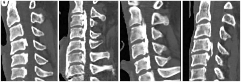
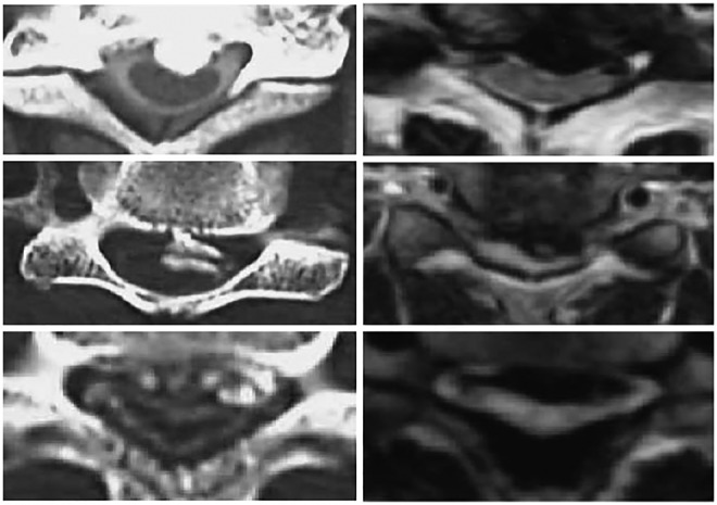
Results: A total of 167 studies were deemed relevant. Five classification systems were developed based on X-ray: the 9-classification system (0.60%); continuous, segmental, mixed, localized or focal, circumscribed and others (92.81%); hook, staple, bridge, and total types (2.40%); distribution of OPLL (2.40%); and K-line classification (4.19%). Six methods were based on computed tomography scans: free-type, contiguous-type, and broken sign (0.60%); hill-, plateau-, square-, mushroom-, irregular-, or round-shaped (5.99%); rectangular, oval, triangular, or pedunculate (1.20%); centralized or laterally deviated (1.80%); plank-, spindle-, or rod-shaped (0.60%); and rule of nine (0.60%). Classification systems based on 3-dimensional computed tomography were bridging and nonbridging (1.20%) and flat, irregular, and localized (0.60%). A single classification system was based on magnetic resonance imaging: triangular, teardrop, or boomerang. Finally, a variation of methods was used to classify OPLL associated with the dura mater (4.19%).
Conclusions: The most common method of classification was that proposed by the Japanese Ministry of Health, Labour and Welfare. Other important methods include K-line (+/-), signs of dural ossification, and patterns of distribution.
Keywords: K-line classification; classification systems; dural ossification; ossification of the posterior longitudinal ligament; reliability.
Conflict of interest statement
Declaration of Conflicting Interests: The author(s) declared no potential conflicts of interest with respect to the research, authorship, and/or publication of this article.
Figures


References
-
- Matsunaga S, Sakou T. Ossification of the posterior longitudinal ligament of the cervical spine: etiology and natural history. Spine (Phila Pa 1976). 2012;37:E309–E314. - PubMed
-
- Stapleton CJ, Pham MH, Attenello FJ, Hsieh PC. Ossification of the posterior longitudinal ligament: genetics and pathophysiology. Neurosurg Focus. 2011;30(3):E6. - PubMed
-
- Nouri A, Tetreault L, Singh A, Karadimas SK, Fehlings MG. Degenerative cervical myelopathy: epidemiology, genetics, and pathogenesis. Spine (Phila Pa 1976). 2015;40:E675–E693. - PubMed
-
- Nakanishi T, Mannen T, Toyokura Y. Asymptomatic ossification of the posterior longitudinal ligament of the cervical spine. Incidence and roentgenographic findings. J Neurol Sci. 1973;19:375–381. - PubMed
-
- Aita I, Ohno A, Amagai H, Hirabayashi H, Hayashi K. Histomorphometric study of iliac bones in cervical myelopathy with ossification of the posterior longitudinal ligament. J Orthop Sci. 1998;3:324–329. - PubMed
Publication types
LinkOut - more resources
Full Text Sources
