Case Reports
. 2018;8(4):485-488.
doi: 10.4314/ovj.v8i4.21. Epub 2018 Dec 27.

Agenesis of hepatic lobes in a dog

Affiliations
Case Reports

Agenesis of hepatic lobes in a dog

Yasuhisa Oishi et al. Open Vet J. 2018.

Abstract

Agenesis of a hepatic lobe is an extremely rare congenital anomaly and only one dog have been reported in veterinary literature. We encountered a dog with this anomaly diagnosed by Computed tomography (CT) and portography. A two-year-old, 6.9-kg female Shih tzu dog was presented with vomiting and anorexia. The dog had no history of abdominal surgery or trauma. Biochemical analysis showed elevated plasmatic liver enzymes. CT revealed the absence of the liver parenchyma and vascular system of the left lobe, quadrate lobe and papillary process of the caudate lobe. A portosystemic shunt was also observed. The liver parenchyma and vascular system of these lobes were not detected under digital subtraction angiography during laparotomy. Furthermore, the liver parenchyma and vascular system of these lobes were not detected even when the remaining liver volume increased two months after treating the shunt vessel. CT proved itself a good option for antemortally diagnosis of hepatic agenesis in a dog.

Keywords: Computed tomography; Dog; Hepatic lobe agenesis; Liver; Portosystemic shunts.


Figures

Fig. 1
Fig. 1
Abdominal radiographs of a 2-year-old female Shih tzu dog. (A): Ventral-dorsal radiograph reveals that gastric silhouette is displaced toward the left abdominal wall. (B): lateral radiograph reveals gastric silhouette parallel with the diaphragm outline.
Fig. 2
Fig. 2
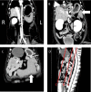
Transverse computed tomography images of a two-year-old female Shih tzu dog before surgical treatment. (A): Dorsal–ventral abdominal radiograph. Three lines reveal each transverse section level. Transverse computed tomography images at levels of (B) the tenth, (C) the eleventh and (D) the twelfth thoracic vertebrae in the portal phase revealed an enlarged bile duct (large arrow: blue) and gallbladder (arrowhead: red) and a shunt vessel (small arrow: yellow) between the caudal vena cava and left gastric vein. No left liver parenchyma was observed at any transverse section levels.
Fig. 3
Fig. 3
Computed tomography multiplanar reconstruction (MPR) images of a two-year-old female Shih tzu dog before surgical treatment. (A): Sagittal MPR image. Three lines reveal each dorsal section level. (B-D): Dorsal MPR images reveal that the greater part of the left diaphragm is attached to the stomach and the remaining liver exists only on the right side of the gallbladder. Large arrow (blue) shows spleen, small arrow (yellow) shows pancreas, large arrowhead (red) showed bile duct and small arrowhead (red) shows the gallbladder. No left liver parenchyma was observed at any dorsal section levels.
Fig. 4
Fig. 4
Portography and three-dimensional computed tomography (3D-CT) images of a two-year-old female Shih tzu dog. (A): Portography after temporary occlusion in the remaining liver. Portal veins of the right medial hepatic lobe (large black arrow), right lateral hepatic lobe (white arrowhead) and caudate process of the caudate lobe (white arrow) were visualized, but no other vessels were observed. Large white arrow shows a gastroduodenal vein. (B): Preoperative 3D-CT image. Preoperative liver volume was 127 cc (18.4 cc/kg). Yellow: abnormal shunt vessel, red: artery, blue: vena cava and purple: portal vein. (C): Postoperative 3D-CT image. Postoperative liver volume was 187 cc (27.1 cc/kg). Gastroduodenal vessels were moved to left after surgery due to liver volume increasing.

References

    1. Buob S, Johnston A.N, Webster C.R. Portal hypertension: pathophysiology, diagnosis, and treatment. J. Vet. Intern. Med. 2011;25:169–186. - PubMed
    1. Doran I.P, Barr F.J, Hotston Moore A, Knowles T.G, Holt P.E. Liver size, bodyweight, and tolerance to acute complete occlusion of congenital extrahepatic portosystemic shunts in dogs. Vet. Surg. 2008;37:656–662. - PubMed
    1. Iannelli A, Facchiano E, Fabiani P, Sejor E, Bernard J.L, Niezar E, Gugenheim J. Agenesis of the right liver: a difficult laparoscopic cholecystectomy. J. Laparoendosc. Adv. Surg. Tech. A. 2005;15:166–169. - PubMed
    1. Inoue T, Ito Y, Matsuzaki Y, Okauchi Y, Kondo H, Horiuchi N, Nakao K, Iwata M. Hypogenesis of right hepatic lobe accompanied by portal hypertension: case report and review of 31 Japanese cases. J. Gastroenterol. 1997;32:836–842. - PubMed
    1. Kakitsubata Y, Nakamura R, Mitsuo H, Suzuki Y, Kakitsubata S, Watanabe K. Absence of the left lobe of the liver: US and CT appearance. Gastrointest. Radiol. 1991;16:323–325. - PubMed

Publication types

LinkOut - more resources