

Primary endometrial large cell neuroendocrine carcinoma with melanocytic differentiation
- PMID: 30775319
- PMCID: PMC6360826
- DOI: 10.4322/acr.2018.041
Primary endometrial large cell neuroendocrine carcinoma with melanocytic differentiation
Abstract
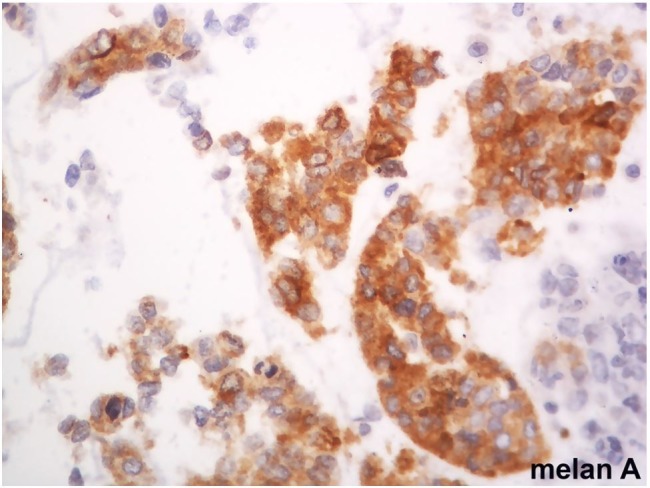
High-grade endometrial carcinomas are aggressive neoplasms of difficult histological classification. Neuroendocrine differentiation in endometrial carcinomas is rare. This is the report of an endometrial large cell neuroendocrine carcinoma with foci of melanocytic differentiation in a 75-year-old woman with abnormal post-menopausal uterine bleeding for 2 years. Two initial biopsies were inconclusive. Histopathological examination of the uterus revealed large cell neuroendocrine carcinoma associated with endometrioid carcinoma and foci of melanocytic differentiation, pT3a (FIGO IIIA). There were metastases in the rectum serosa and lungs. After 8 months of diagnosis and surgical treatment, the patient is on chemotherapy and radiotherapy. We highlight the morphological characteristics and criteria that allow the definitive anatomopathological diagnosis, including immunohistochemical markers used to identify the cell types present in this unprecedented association.
Keywords: Carcinoma, Large Cell; Carcinoma, Neuroendocrine; Endometrium; Immunohistochemistry; Melanocytes.
Conflict of interest statement
Conflict of interest: None
Figures


References
-
- Kumar V, Abbas AK, Aster JC, editors. Robbins and Cotran pathologic basis of disease. 9th ed. Philadelphia: Elsevier/Saunders; 2015.
-
- Kurman RJ, Ellenson LH, Ronnett BM, editors. Blaunstein’s pathology of the female genital tract. 6th ed. New York: Springer; 2011. 10.1007/978-1-4419-0489-8. - DOI
-
- Instituto Nacional de Câncer José Alencar Gomes da Silva Coordenação de Prevenção e Vigilância Estimativa 2018: incidência de câncer no Brasil [Internet]. Rio de Janeiro: INCA; 2017. [cited 2018 May 17]. Available from: http://www.inca.gov.br/estimativa/2018/
