Case Reports
. 2018 Sep 26;8(4):e2018043.
doi: 10.4322/acr.2018.043. eCollection 2018 Oct-Dec.

Malignant ameloblastoma: a challenging diagnosis

Affiliations
Case Reports

Malignant ameloblastoma: a challenging diagnosis

Ayodeji Salami et al. Autops Case Rep. .

Abstract

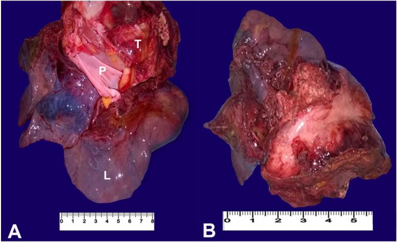
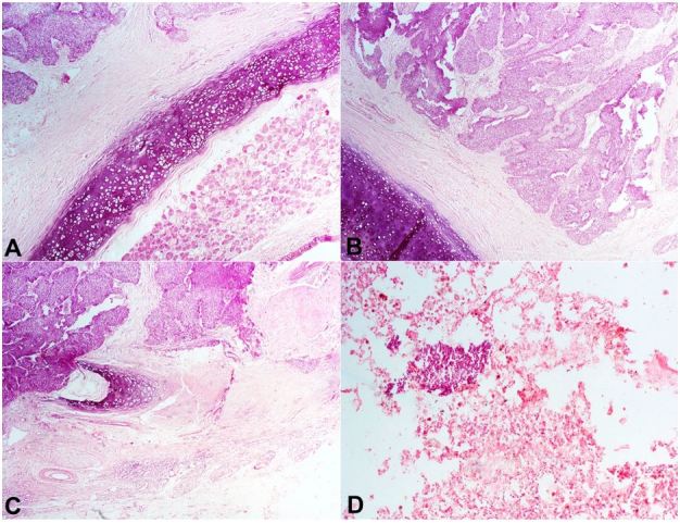
Ameloblastoma is an uncommon and locally aggressive, benign, odontogenic tumor, with local recurrence when not adequately excised. A rare variant of this neoplasm with the benign features but accompanied with metastases has been described. This rare variant is malignant ameloblastoma and is known to have a poor prognosis. We present the case of a young woman who had recurrent mandibular tumors, which were resected twice and histologically reported as ameloblastoma. Four years later, she presented with pulmonary metastasis and atelectasis. A review of the literature on this very rare neoplasm was also performed.

Keywords: Ameloblastoma; Mediastinum; Neoplasm Metastasis; Pulmonary Atelectasis.


Figures

Figure 1
Figure 1. Gross view of the neoplasm. A – The tumor (T) attached to the left lung (L) and the parietal pericardium (P); B – The cut surface of the tumor shows a greyish-white mass attached to the lung surface.
Figure 2
Figure 2. Photomicrography of the tumor. A – Nests and trabeculae of malignant cells show peripheral palisading of darkly staining cells with loose cells in the center (H&E, 250X); B and C – High power view showing stellate cells having a reticular appearance, and palisading columnar to polygonal cells at the periphery (H&E, 400X).
Figure 3
Figure 3. Photomicrography of the tumor. A, B and C – Infiltration of the trachea and bronchi by the malignant cells (H&E, 250X); D – A nest of tumor cells within the lung parenchyma (H&E, 250X).

References

    1. McClary AC, West RB, McClary AC, et al. . Ameloblastoma: a clinical review and trends in management. Eur Arch Otorhinolaryngol. 2016;273(7):1649-61. 10.1007/s00405-015-3631-8. - DOI - PubMed
    1. Jayaraj G, Sherlin HJ, Ramani P, et al. . Metastasizing ameloblastoma - a perennial pathological enigma? Report of a case and review of literature. J Craniomaxillofac Surg. 2014;42(6):772-9. 10.1016/j.jcms.2013.11.009. - DOI - PubMed
    1. Ajagbe HA, Daramola JO. Ameloblastoma: a survey of 199 cases in the University of College Hospital, Ibadan, Nigeria. J Natl Med Assoc. 1987;79(3):324-7. - PMC - PubMed
    1. Ladeinde AL, Ogunlewe MO, Bamgbose BO, et al. . Ameloblastoma: analysis of 207 cases in a Nigerian teaching hospital. Quintessence Int. 2006;37(1):69-74. - PubMed
    1. Kodati S, Majumdar S, Uppala D, Namana M. Ameloblastic carcinoma: a report of three cases. J Clin Diagn Res. 2016;10(10):23-5. - PMC - PubMed

Publication types