

Fracture resistance of simulated immature teeth treated with a regenerative endodontic protocol
- PMID: 30775411
- PMCID: PMC6366405
- DOI: 10.1080/23337931.2019.1570822
Fracture resistance of simulated immature teeth treated with a regenerative endodontic protocol
Abstract
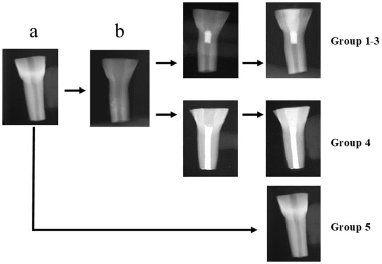
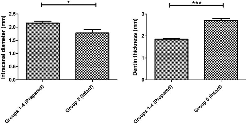
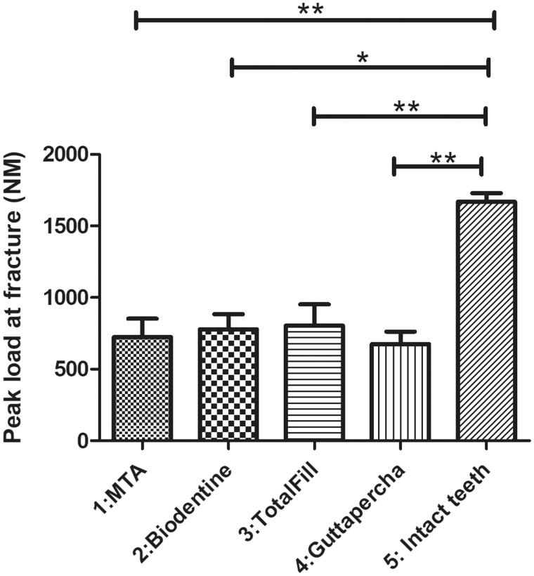
This study aims to evaluate fracture resistance of simulated immature teeth after treatment with regenerative endodontic procedure (REP) using tricalcium silicate cements (TSCs) as cervical plugs. Bovine incisors were sectioned to standard crown/root ratio. Pulp tissue was removed and canals were enlarged to a standardized diameter. Teeth were then treated with a REP protocol consisting of NaOCl and EDTA irrigation, intracanal medication with triple-antibiotic paste for 14 days followed by a TSC cervical seal and composite restoration. Teeth were divided into groups according to the material used; Mineral-Trioxide-Aggregate (MTA), Biodentine, TotalFill. Teeth filled with guttapercha (GP) and intact teeth served as controls. All teeth subjected to an increasing compressive force (rate of 0.05 mm/s at a 45° angle to the long axis of the tooth) until fracture. All treated teeth exhibited significantly lower resistance to fracture compared to the intact teeth but no difference was found between the TSC groups (Kruskal-Wallis, Dunn's multiple comparison, p < .05). TSCs applied at the cervical area of simulated immature teeth treated with REP did not reinforce fracture resistance.
Keywords: Biodentine; Fracture resistance; MTA; TotalFill; bovine teeth.
Figures
References
-
- Cvek M. Prognosis of luxated non-vital maxillary incisors treated with calcium hydroxide and filled with gutta-percha. A retrospective clinical study. Dent Traumatol. 1992;8:45–55. - PubMed
-
- Glendor U. Epidemiology of traumatic dental injuries-a 12 year review of the literature. Dent Traumatol. 2008;24:603–611. - PubMed
-
- Andreasen JO, Munksgaard EC, Bakland LK. Comparison of fracture resistance in root canals of immature sheep teeth after filling with calcium hydroxide or MTA. Dent Traumatol. 2006;22:154–156. - PubMed
-
- Sawyer AN, Nikonov SY, Pancio AK, et al. Effects of calcium silicate-based materials on the flexural properties of dentin. J Endod. 2012;38:680–683. - PubMed
-
- Bortoluzzi EA, Souza EM, Reis JM, et al. Fracture strength of bovine incisors after intra-radicular treatment with MTA in an experimental immature tooth model. Int Endod J. 2007;40:684–691. - PubMed
LinkOut - more resources
Full Text Sources