. 2019 Mar 12;58(10):1354-1362.
doi: 10.1021/acs.biochem.8b01290. Epub 2019 Feb 22.

Role of Backbone Dynamics in Modulating the Interactions of Disordered Ligands with the TAZ1 Domain of the CREB-Binding Protein

Affiliations

Role of Backbone Dynamics in Modulating the Interactions of Disordered Ligands with the TAZ1 Domain of the CREB-Binding Protein

Rebecca B Berlow et al. Biochemistry. .

Abstract

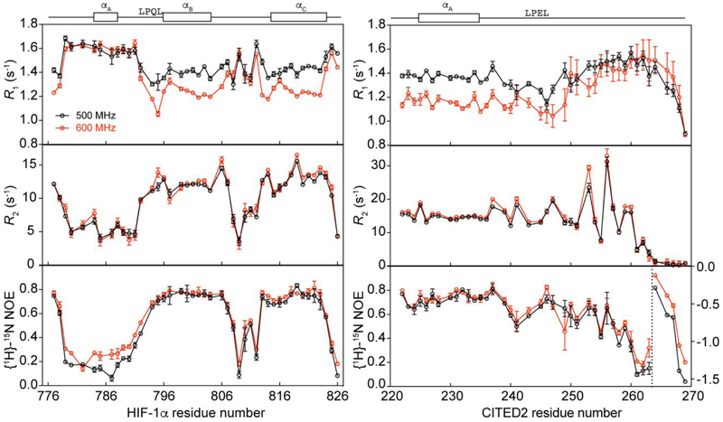
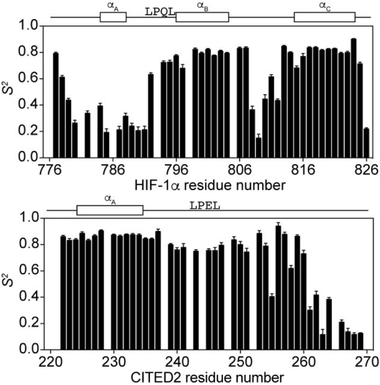
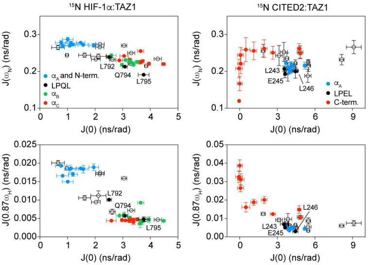
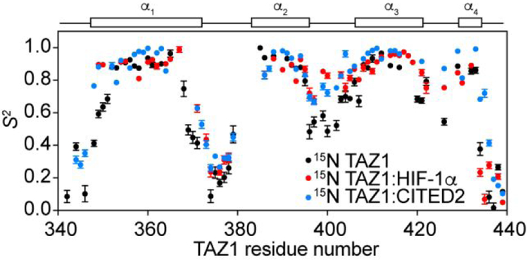
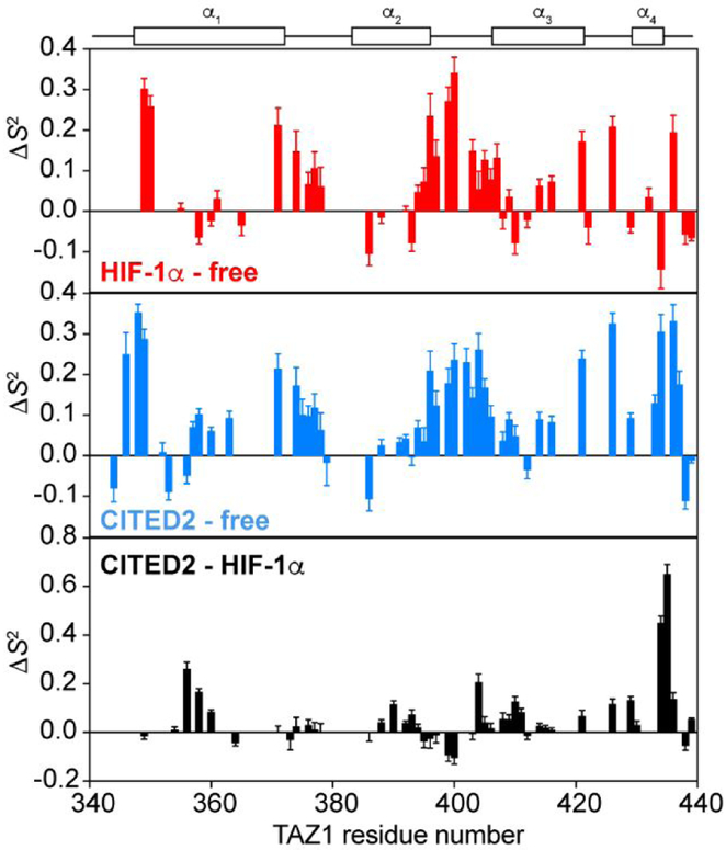
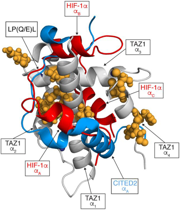
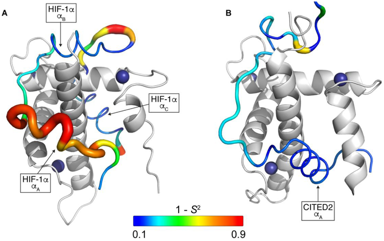
The intrinsically disordered transactivation domains of HIF-1α and CITED2 compete for binding of the TAZ1 domain of the CREB-binding protein by a unidirectional allosteric mechanism involving direct competition for shared binding sites, ternary complex formation, and TAZ1 conformational changes. To gain insight into the mechanism by which CITED2 displaces HIF-1α from TAZ1, we used nuclear magnetic resonance spin relaxation methods to obtain an atomic-level description of the picosecond to nanosecond backbone dynamics that contribute to TAZ1 binding and competition. We show that HIF-1α and CITED2 adopt different dynamics in their complexes with TAZ1, with flexibility observed for HIF-1α in regions that would maintain accessibility for CITED2 to bind to TAZ1 and facilitate subsequent HIF-1α dissociation. In contrast, critical regions of CITED2 adopt a rigid structure in its complex with TAZ1, minimizing the ability of HIF-1α to compete for binding. We also find that TAZ1, previously thought to be a rigid scaffold for binding of disordered protein ligands, displays altered backbone dynamics in its various bound states. TAZ1 is more rigid in its CITED2-bound state than in its free state or in complex with HIF-1α, with increased rigidity observed not only in the CITED2 binding site but also in regions of TAZ1 that undergo conformational changes between the HIF-1α- and CITED2-bound structures. Taken together, these data suggest that backbone dynamics in TAZ1, as well as in the HIF-1α and CITED2 ligands, play a role in modulating the occupancy of TAZ1 and highlight the importance of characterizing both binding partners in molecular interactions.


Figures

Figure 1.
Figure 1.
NMR spin relaxation data for 15N HIF-1α:TAZ1 and 15N CITED2:TAZ1 complexes. 15N R1, 15N R2, and {1H}-15N NOE data collected at 500 MHz (black) and 600 MHz (red) are shown for 15N HIF-1α:TAZ1 (left) and 15N CITED2:TAZ1 (right). The secondary structures formed by HIF-1α (left) and CITED2 (right) in complex with TAZ1 and the conserved LP(Q/E)L motif are shown above the graphs for reference. {1H}-15N NOE data for CITED2 residues 264-269 (to the right of the dashed vertical line) are plotted on the right y-axis.
Figure 2.
Figure 2.
Backbone amide order parameters (S2) for 15N HIF-1α:TAZ1 and 15N CITED2:TAZ1 complexes. S2 values obtained from Model-Free analysis of the NMR spin relaxation data are shown for 15N HIF-1α:TAZ1 (top) and 15N CITED2:TAZ1 (bottom).The secondary structures formed by HIF-1α (top) and CITED2 (bottom) in complex with TAZ1 and the conserved LP(Q/E)L motif are shown above the graphs for reference.
Figure 3.
Figure 3.
Evidence for dynamic clustering within binding motifs in 15N HIF-1α:TAZ1 and 15N CITED2:TAZ1. The spectral densities J(ωN) (top) and J(0.87ωH) (bottom) are plotted as a function of J(0) for 15N HIF-1α:TAZ1 (left) and 15N CITED2:TAZ1 (right) for data collected at 600 MHz. Data corresponding to discrete binding motifs of HIF-1α and CITED2 are shown in the colors indicated in the legends. Data points corresponding to the residues in the conserved LP(Q/E)L motif are labeled.
Figure 4.
Figure 4.
Backbone amide order parameters (S2) for 15N TAZ1,15N TAZ1:HIF-1α and 15N TAZ1:CITED2 complexes. S2 values obtained from Model-Free analysis of the NMR spin relaxation data are shown for 15N TAZ1 (black), 15N TAZ1:HIF-1α (red), and 15N TAZ1:CITED2 (blue). The helical regions of TAZ1 are shown above the graph for reference.
Figure 5.
Figure 5.
Changes in 15N TAZ1 backbone amide order parameters upon HIF-1α or CITED2 binding. Differences in S2 values (ΔS2) obtained from Model-Free analysis are shown for 15N TAZ1:HIF-1α compared to free 15N TAZ1 (top), 15N TAZ1:CITED2 compared to free 15N TAZ1 (middle), and 15N TAZ1:CITED2 compared to 15N TAZ1:HIF-1α (bottom). The helical regions of TAZ1 are shown above the graphs for reference.
Figure 6.
Figure 6.
Mapping changes in 15N TAZ1 backbone amide order parameters between the 15N TAZ1:HIF-1α and 15N TAZ1:CITED2 complexes. TAZ1 is shown in grey, HIF-1α is shown in red, and CITED2 is shown in blue. Zinc atoms are shown as dark blue spheres. Residues with ΔS2 greater than 0.1 are shown as orange spheres. The ΔS2 values were calculated as S2CITED2 - S2HIF1α. The secondary structural elements of HIF-1α and CITED2, the conserved LP(Q/E)L motif, and the TAZ1 helices are labeled for reference. The disordered C-terminal tail of CITED2 (residues 260-269) is not shown.
Figure 7.
Figure 7.
Backbone dynamics for HIF-1α and CITED2 in their binary complexes with TAZ1. The ribbon width of the HIF-1α (A) and CITED2 (B) transactivation domain peptides is scaled by 1-S2; the backbone color gradient ranges from blue (1-S2 = 0.1) to red (1- S2 = 0.9). TAZ1 is shown in the cartoon representation in grey. Zinc atoms are shown as dark blue spheres. The disordered C-terminal tail of CITED2 (residues 260-269) is not shown. The secondary structural elements of HIF-1α and CITED2 are labeled for reference.

References

    1. Wright PE, and Dyson HJ (2015) Intrinsically disordered proteins in cellular signalling and regulation, Nat Rev Mol Cell Biol 16, 18–29. - PMC - PubMed
    1. van der Lee R, Buljan M, Lang B, Weatheritt RJ, Daughdrill GW, Dunker AK, Fuxreiter M, Gough J, Gsponer J, Jones DT, Kim PM, Kriwacki RW, Oldfield CJ, Pappu RV, Tompa P, Uversky VN, Wright PE, and Babu MM (2014) Classification of intrinsically disordered regions and proteins, Chemical reviews 114, 6589–6631. - PMC - PubMed
    1. Modell AE, Blosser SL, and Arora PS (2016) Systematic Targeting of Protein-Protein Interactions, Trends Pharmacol Sci 37, 702–713. - PMC - PubMed
    1. Sammak S, and Zinzalla G. (2015) Targeting protein-protein interactions (PPIs) of transcription factors: Challenges of intrinsically disordered proteins (IDPs) and regions (IDRs), Progress in biophysics and molecular biology 119, 41–46. - PubMed
    1. Tsafou K, Tiwari PB, Forman-Kay JD, Metallo SJ, and Toretsky JA (2018) Targeting Intrinsically Disordered Transcription Factors: Changing the Paradigm, J Mol Biol 430, 2321–2341. - PubMed

Publication types

MeSH terms