Review
. 2019 Apr 1;129(4):1463-1474.
doi: 10.1172/JCI124608. Epub 2019 Feb 18.

Epithelial barrier repair and prevention of allergy

Affiliations
Review

Epithelial barrier repair and prevention of allergy

Elena Goleva et al. J Clin Invest. .

Abstract

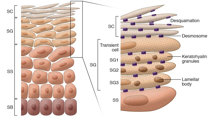
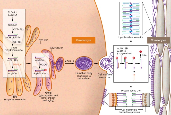
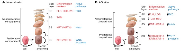
Allergic diseases have in common a dysfunctional epithelial barrier, which allows the penetration of allergens and microbes, leading to the release of type 2 cytokines that drive allergic inflammation. The accessibility of skin, compared with lung or gastrointestinal tissue, has facilitated detailed investigations into mechanisms underlying epithelial barrier dysfunction in atopic dermatitis (AD). This Review describes the formation of the skin barrier and analyzes the link between altered skin barrier formation and the pathogenesis of AD. The keratinocyte differentiation process is under tight regulation. During epidermal differentiation, keratinocytes sequentially switch gene expression programs, resulting in terminal differentiation and the formation of a mature stratum corneum, which is essential for the skin to prevent allergen or microbial invasion. Abnormalities in keratinocyte differentiation in AD skin result in hyperproliferation of the basal layer of epidermis, inhibition of markers of terminal differentiation, and barrier lipid abnormalities, compromising skin barrier and antimicrobial function. There is also compelling evidence for epithelial dysregulation in asthma, food allergy, eosinophilic esophagitis, and allergic rhinosinusitis. This Review examines current epithelial barrier repair strategies as an approach for allergy prevention or intervention.


Conflict of interest statement

Conflict of interest: The authors have declared that no conflict of interest exists.

Figures

Figure 1
Figure 1. Structure of human epidermis.
Human epidermis is composed of SB, SS, SG, and SC. In SC, corneocytes (flattened and denucleated keratinocytes) and intercellular lipids released from lamellar bodies form the “brick and mortar” structures. The cornified envelope, a highly cross-linked layer of insoluble proteins, forms under the corneocyte cell membrane, anchored by extracellular lipids. Components of the cornified envelope (keratohyalin granules, a source of filaggrin) and lamellar bodies (containing lipids, lipid-processing enzymes, corneodesmosin, proteases, and protease inhibitors) are formed in SG. The surface of the SC is shed off by degradation of corneodesmosomes via the activity of several proteases.
Figure 2
Figure 2. Lamellar body formation and generation of acylceramides and protein-bound ceramides in human epidermis.
The enzymes and reactions responsible for creating acylceramides and protein-bound ceramides are shown. The schematics represent acylceramide production, lamellar body assembly and secretion in SG, and formation of the protein-bound ceramides in SC. Acylceramides are formed in the ER and secreted through the Golgi apparatus. ω-OH protein-bound ceramides are formed at the cell membrane. Sequential actions of the fatty acid elongases ELOVL1 and ELOVL4 generate ULCFAs of up to C26 and C28 carbon-chain-lengths, respectively. The cytochrome P450 enzyme CYP4F22 then ω-hydroxylates these ULCFAs, generating ω-OH ULCFAs. Next, the ceramide synthase CERS3 uses ω-OH ULCFAs for ω-OH ceramide synthesis. Finally, the transacylase PNPLA1 forms an ester linkage between the 18:2n-6 fatty acid taken from triglycerides and the ω-OH group of ω-OH ceramide to create an acylceramide. Each enzyme involved in acylceramide production is localized in the ER, indicating that acylceramide production takes place there. Once produced, the UDP-glucose ceramide glucosyltransferase UGCG glycosylates acylceramides in the Golgi apparatus, followed by ABCA12-mediated transport into lamellar bodies. In the course of protein-bound ceramide production, the 18:2n-6 fatty acid portions of acylceramides are subjected to peroxidation by the lipoxygenases ALOX12B and ALOXE3, followed by deglycosylation by β-glucosylceramidase (GBA). Transglutaminase then cross-links the exposed ω-OH group with cornified envelope proteins such as involucrin, envoplakin, and periplakin. Cer, ceramide; GlcCer, glucosylceramide; G, glycosyl group.
Figure 3
Figure 3. Epidermal differentiation pattern in normal and AD skin.
(A and B) Epidermal differentiation pattern in normal (A) and AD (B) skin. The keratinocyte differentiation process is under tight regulation. Cells proliferate in the basal layer of the epidermis. As basal layer keratinocytes detach from the basement membrane and migrate into the first suprabasal layer in the spinous layer, they irreversibly exit the cell cycle and switch from KRT5/KRT14 to KRT1/KRT10 production. During epidermal differentiation, keratinocytes sequentially switch gene expression programs and express the granular layer differentiation markers FLG, LOR, and TGM1. The Wnt/β-catenin pathway is active in the proliferating epidermis, whereas keratinocyte differentiation in the spinous layer is under the control of the Notch pathway. Changes in extracellular Ca2+ and lipid metabolism trigger protein kinase C (PKC) pathway activation and regulate the transcription of FLG, LOR, IVL, and TGM1. In AD skin, abnormalities in the differentiation of keratinocytes result in hyperproliferation of the basal layer, reduction of the spinous layer, and inhibition of markers of terminal differentiation, all of which compromise skin barrier and antimicrobial function.

References

    1. Holgate ST, Wenzel S, Postma DS, Weiss ST, Renz H, Sly PD. Asthma. Nat Rev Dis Primers. 2015;1:15025. - PMC - PubMed
    1. Renz H, et al. Food allergy. Nat Rev Dis Primers. 2018;4:17098. - PubMed
    1. Weidinger S, Beck LA, Bieber T, Kabashima K, Irvine AD. Atopic dermatitis. Nat Rev Dis Primers. 2018;4(1):1. doi: 10.1038/s41572-018-0001-z. - DOI - PubMed
    1. Brunner PM, Guttman-Yassky E, Leung DY. The immunology of atopic dermatitis and its reversibility with broad-spectrum and targeted therapies. J Allergy Clin Immunol. 2017;139(4S):S65–S76. - PMC - PubMed
    1. Yu W, Freeland DMH, Nadeau KC. Food allergy: immune mechanisms, diagnosis and immunotherapy. Nat Rev Immunol. 2016;16(12):751–765. doi: 10.1038/nri.2016.111. - DOI - PMC - PubMed

Publication types