. 2019 May 1;76(5):580-587.
doi: 10.1001/jamaneurol.2018.4921.

Assessing Spinal Cerebrospinal Fluid Leaks in Spontaneous Intracranial Hypotension With a Scoring System Based on Brain Magnetic Resonance Imaging Findings

Affiliations

Assessing Spinal Cerebrospinal Fluid Leaks in Spontaneous Intracranial Hypotension With a Scoring System Based on Brain Magnetic Resonance Imaging Findings

Tomas Dobrocky et al. JAMA Neurol. .

Abstract

Importance: Various signs may be observed on brain magnetic resonance imaging (MRI) in patients with spontaneous intracranial hypotension (SIH). However, the lack of a classification system integrating these findings limits decision making in clinical practice.

Objective: To develop a probability score based on the most relevant brain MRI findings to assess the likelihood of an underlying spinal cerebrospinal fluid (CSF) leak in patients with SIH.

Design, setting, and participants: This case-control study in consecutive patients investigated for SIH was conducted at a single hospital department from February 2013 to October 2017. Patients with missing brain MRI data were excluded. Three blinded readers retrospectively reviewed the brain MRI scans of patients with SIH and a spinal CSF leak, patients with orthostatic headache without a CSF leak, and healthy control participants, evaluating 9 quantitative and 7 qualitative signs. A predictive diagnostic score based on multivariable backward logistic regression analysis was then derived. Its performance was validated internally in a prospective cohort of patients who had clinical suspicion for SIH.

Main outcomes and measures: Likelihood of a spinal CSF leak based on the proposed diagnostic score.

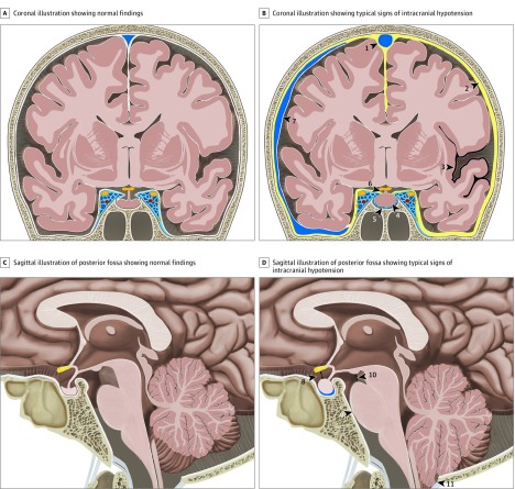
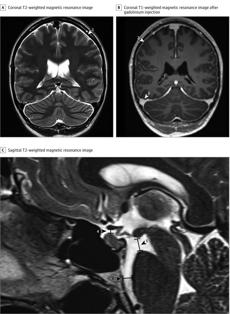
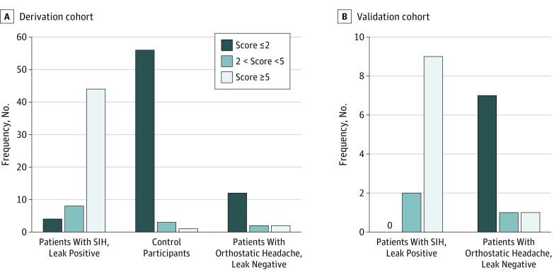
Results: A total of 152 participants (101 female [66.4%]; mean [SD] age, 46.1 [14.3] years) were studied. These included 56 with SIH and a spinal CSF leak, 16 with orthostatic headache without a CSF leak, 60 control participants, and 20 patients in the validation cohort. Six imaging findings were included in the final scoring system. Three were weighted as major (2 points each): pachymeningeal enhancement, engorgement of venous sinus, and effacement of the suprasellar cistern of 4.0 mm or less. Three were considered minor (1 point each): subdural fluid collection, effacement of the prepontine cistern of 5.0 mm or less, and mamillopontine distance of 6.5 mm or less. Patients were classified into groups at low, intermediate, or high probability of having a spinal CSF leak, with total scores of 2 points or fewer, 3 to 4 points, and 5 points or more, respectively, on a scale of 9 points. The discriminatory ability of the proposed score could be demonstrated in the validation cohort.

Conclusions and relevance: This 3-tier predictive scoring system is based on the 6 most relevant brain MRI findings and allows assessment of the likelihood (low, intermediate, or high) of a positive spinal imaging result in patients with SIH. It may be useful in identifying patients with SIH who are leak positive and in whom further invasive myelographic examinations are warranted before considering targeted therapy.


Conflict of interest statement

Conflict of Interest Disclosures: Dr Mossiman receives Swiss National Science Foundation grants for research on brain aneurysms. Dr Gralla reports acting as a global principal investigator of Solitaire FR Thrombectomy for Acute Revascularization study; a clinical events committee member of the Prospective, Multicenter, Observational, Single-Arm European Registry on the ACE Reperfusion Catheters and the Penumbra System in the Treatment of Acute Ischemic Stroke (PROMISE) study (Penumbra); and the principal investigator for the Solitaire with the Intention for Thrombectomy (SWIFT DIRECT) study (Medtronic). Dr Gralla also reports serving as a consultant for Medtronic and receiving Swiss National Science Foundation grants for magnetic resonance imaging in stroke. Dr Beck reports acting as a global principal investigator of Swiss Trial of Decompressive Craniectomy versus Best Medical Treatment of Spontaneous Supratentorial Intracerebral Hemorrhage (SWITCH) and To Scan or Not to Scan (TOSCAN) studies and has receiving a Swiss National Science Foundation grant for ultrasound perfusion imaging. No other disclosures were reported.

Figures

Figure 1.
Figure 1.. Illustration of Typical Findings on Brain Magnetic Resonance Imaging in Intracranial Hypotension
A, Coronal illustration of the brain demonstrating normal findings. B, Coronal illustration of the brain with typical findings in a patient with a spinal cerebrospinal fluid leak with venous engorgement of the superior sagittal sinus (arrowhead 1), pachymeningeal enhancement (arrowhead 2), superficial siderosis (arrowhead 3), enlarged pituitary gland (arrowhead 4), prominent intercavernous sinus (arrowhead 5), effaced suprasellar cistern (arrowhead 6), and subdural fluid collection (arrowhead 7). C, Sagittal illustration of the posterior fossa demonstrating normal findings. D, Sagittal illustration of the posterior fossa with typical findings in patients with a spinal cerebrospinal fluid leak with effaced suprasellar cistern (arrowhead 8; pathologic ≤4 mm), effacement of the prepontine cistern (arrowhead 9; pathologic ≤5 mm), decreased mamillopontine distance (arrowhead 10; pathologic ≤6.5 mm), and low-lying cerebellar tonsils (arrowhead 11).
Figure 2.
Figure 2.. Imaging Findings Included in the Final Diagnostic Score
A, Coronal T2-weighted magnetic resonance imaging (MRI) with subdural effusion (white arrowhead 1). B, Coronal T1-weighted MRI after gadolinium injection of the same patient, with dural enhancement (white arrowhead 2) and venous engorgement (white arrowhead 3). C, Sagittal T2-weighted MRI with effaced suprasellar cistern (black arrowhead 4; pathologic ≤4 mm), decreased mamillopontine distance (black arrowhead 5; pathologic ≤6.5 mm), and effaced prepontine cistern (black arrowhead 6; pathologic ≤5 mm).
Figure 3.
Figure 3.. Predictive Model Scores
A, Scores for individuals in the derivation cohort: patients with spontaneous intracranial hypotension (SIH) who are leak positive, healthy control participants, and patients with orthostatic headache without a cerebrospinal fluid leak. B, Scores for individuals in the internal validation cohort including patients with spontaneous intracranial hypotension and a myelographically confirmed leak and patients presenting with orthostatic headache without a cerebrospinal fluid leak.

References

    1. Vincent M, Wang S; Headache Classification Committee of the International Headache Society . The international classification of headache disorders, 3rd edition. Cephalalgia. 2018;38(1):1-211. doi:10.1177/0333102417738202 - DOI - PubMed
    1. Schievink WI. Spontaneous spinal cerebrospinal fluid leaks and ongoing investigations in this area. JAMA. 2006;295(19):2286-2296. doi:10.1001/jama.295.19.2286 - DOI - PubMed
    1. Fichtner J, Ulrich CT, Fung C, et al. . Management of spontaneous intracranial hypotension—transorbital ultrasound as discriminator. J Neurol Neurosurg Psychiatry. 2016;87(6):650-655. doi:10.1136/jnnp-2015-310853 - DOI - PMC - PubMed
    1. Beck J, Ulrich CT, Fung C, et al. . Diskogenic microspurs as a major cause of intractable spontaneous intracranial hypotension. Neurology. 2016;87(12):1220-1226. doi:10.1212/WNL.0000000000003122 - DOI - PubMed
    1. Beck J, Fung C, Ulrich CT, et al. . Cerebrospinal fluid outflow resistance as a diagnostic marker of spontaneous cerebrospinal fluid leakage. J Neurosurg Spine. 2017;27(2):227-234. doi:10.3171/2017.1.SPINE16548 - DOI - PubMed

MeSH terms