. 2019 Mar 14;62(5):2598-2617.
doi: 10.1021/acs.jmedchem.8b01927. Epub 2019 Mar 5.

Epidermal Growth Factor Receptor-Targeted Multifunctional Photosensitizers for Bladder Cancer Imaging and Photodynamic Therapy

Affiliations

Epidermal Growth Factor Receptor-Targeted Multifunctional Photosensitizers for Bladder Cancer Imaging and Photodynamic Therapy

Ravindra R Cheruku et al. J Med Chem. .

Abstract

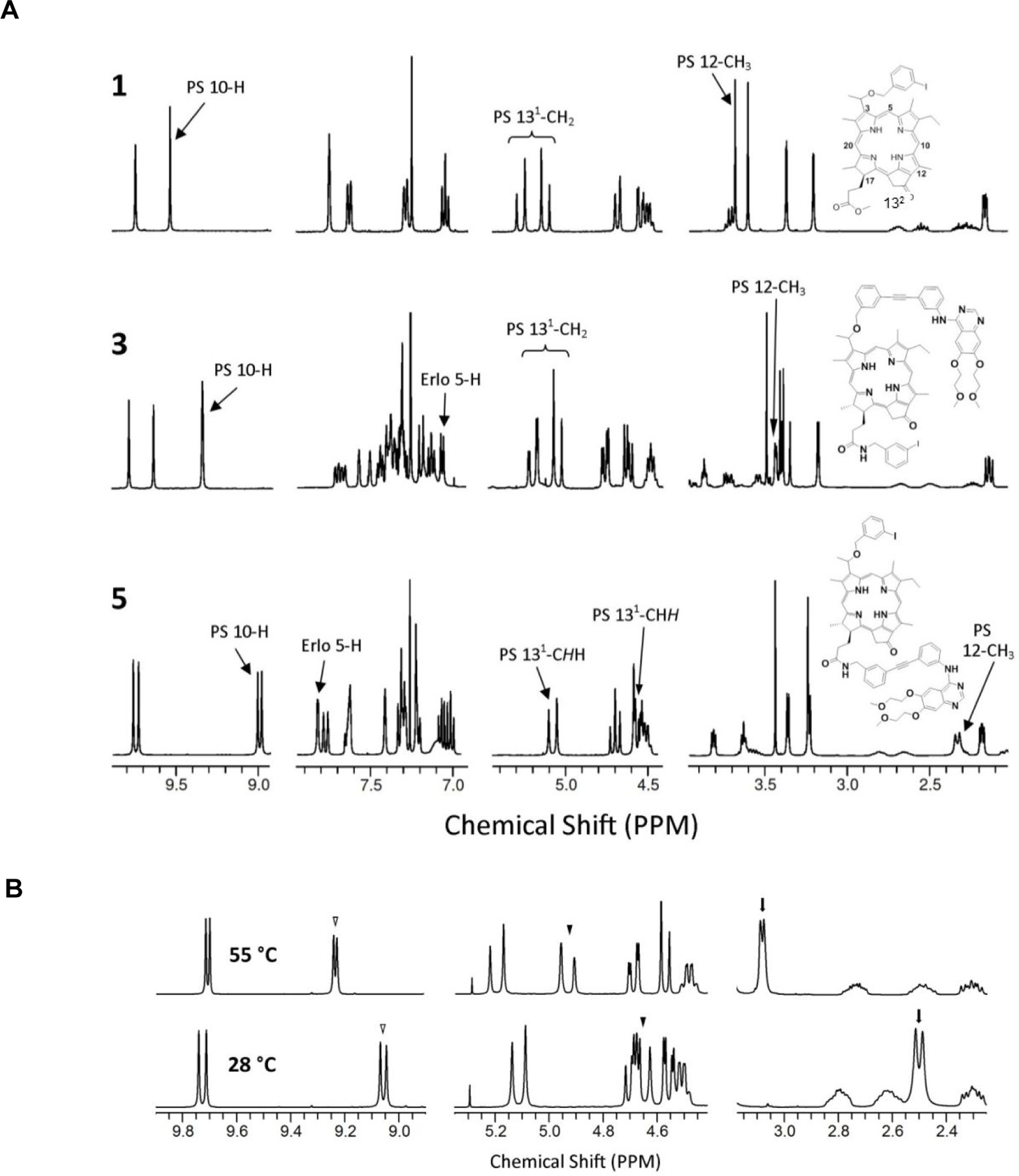
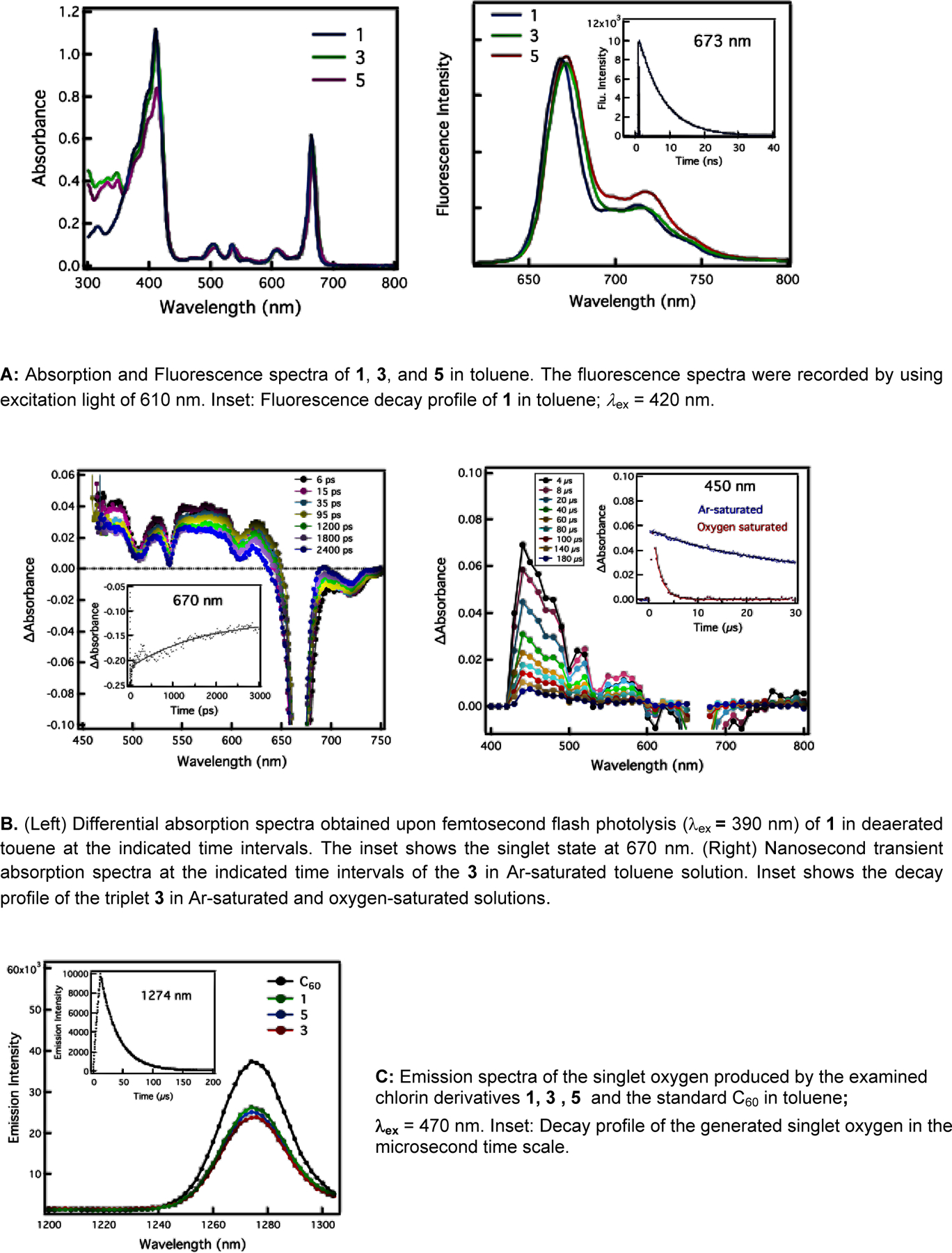
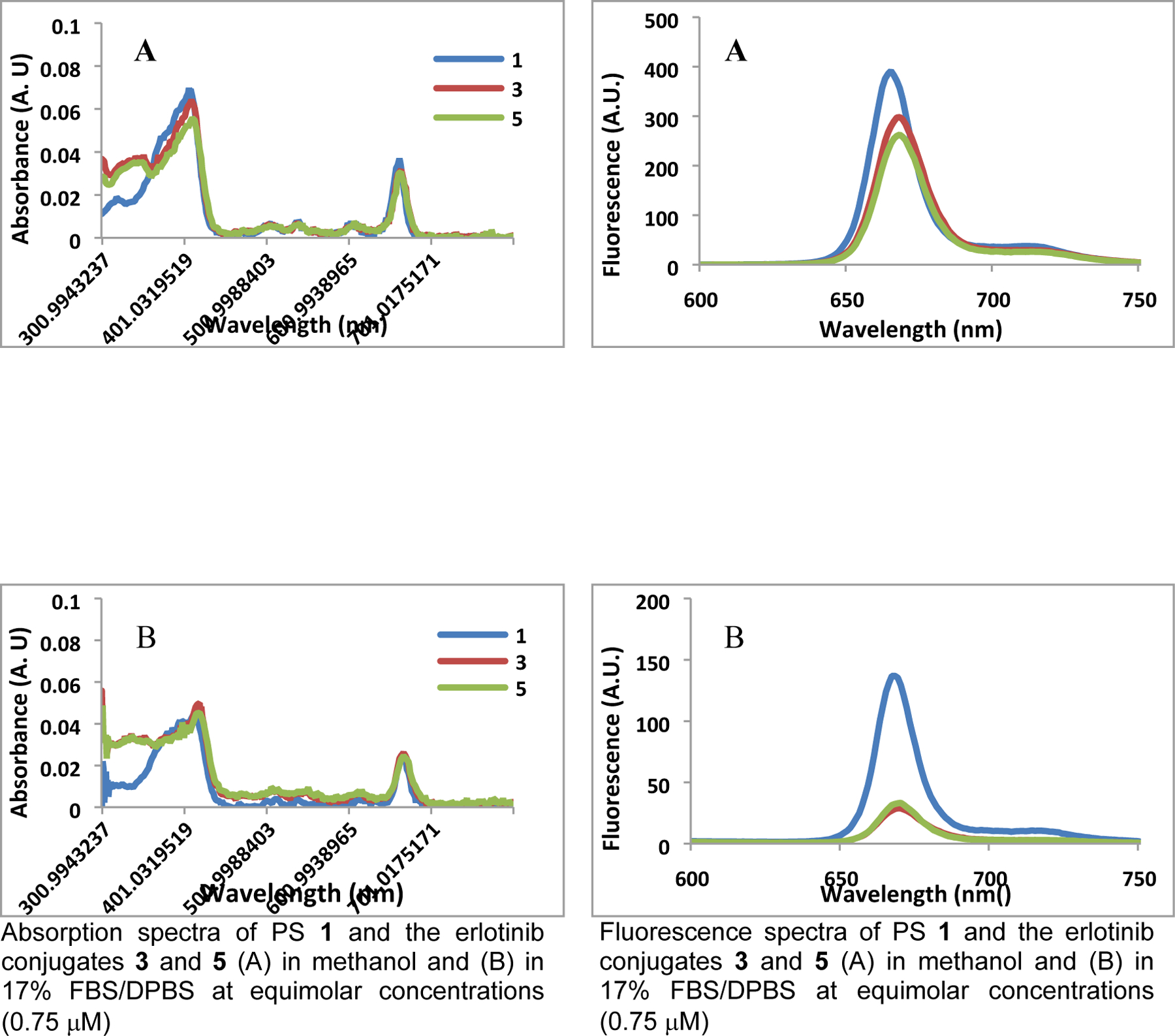
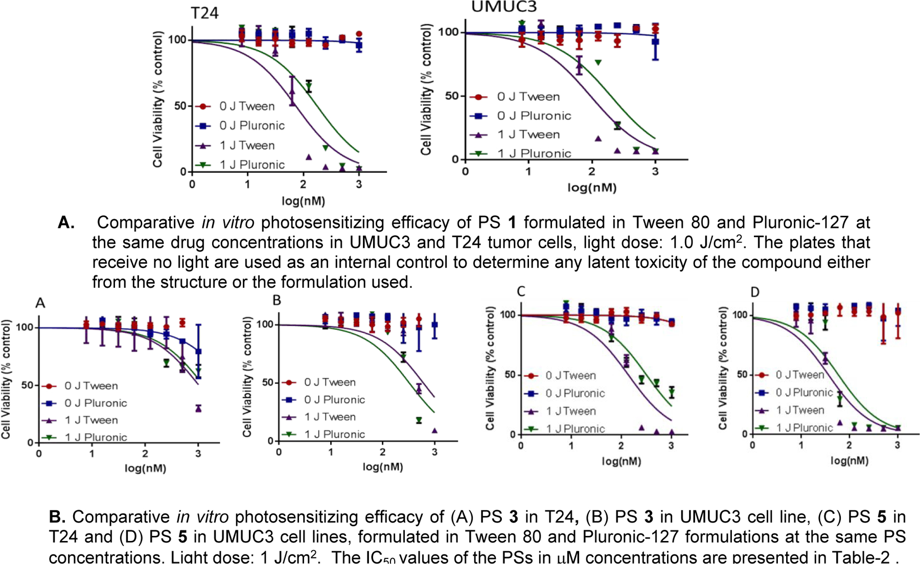
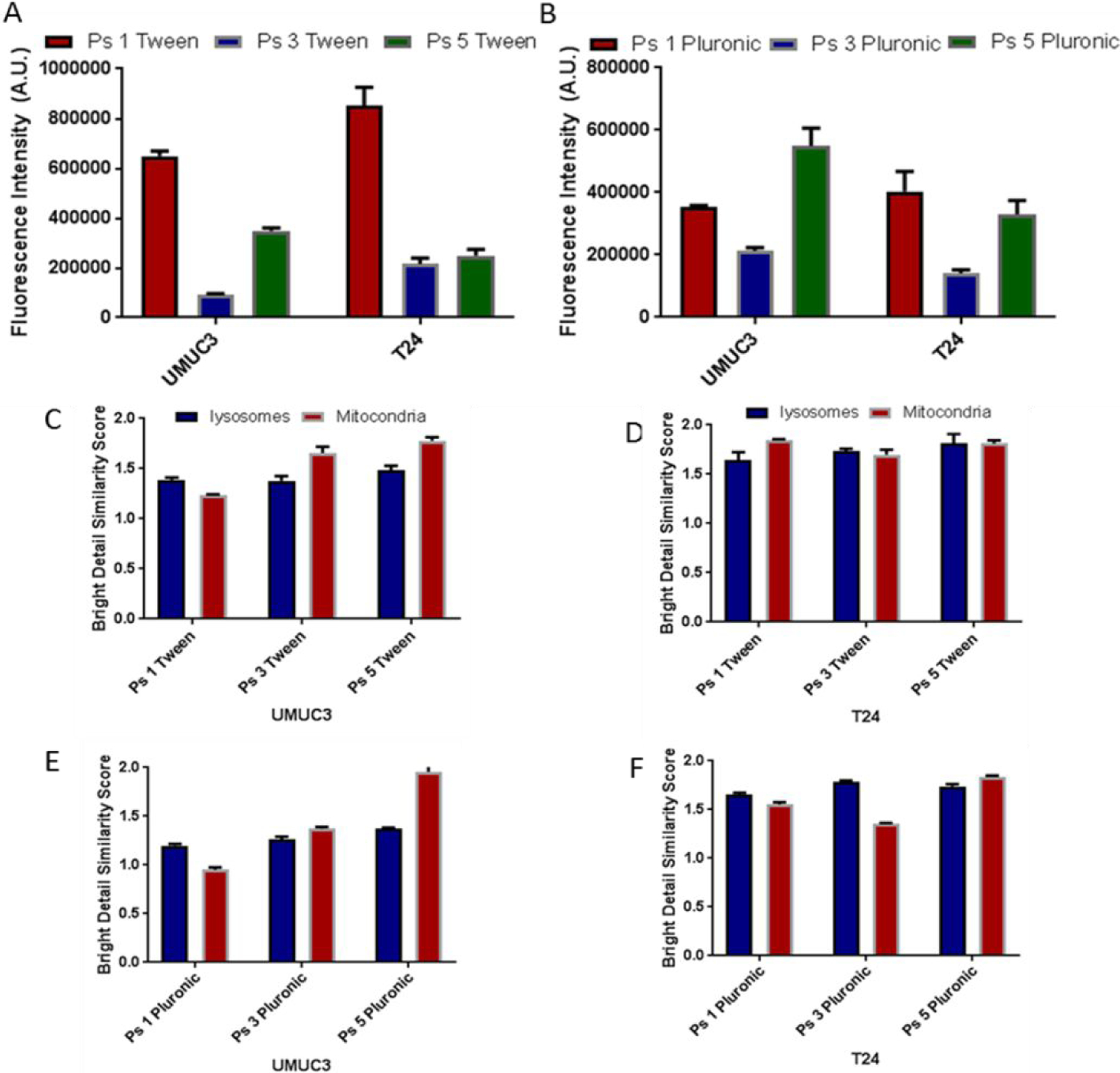
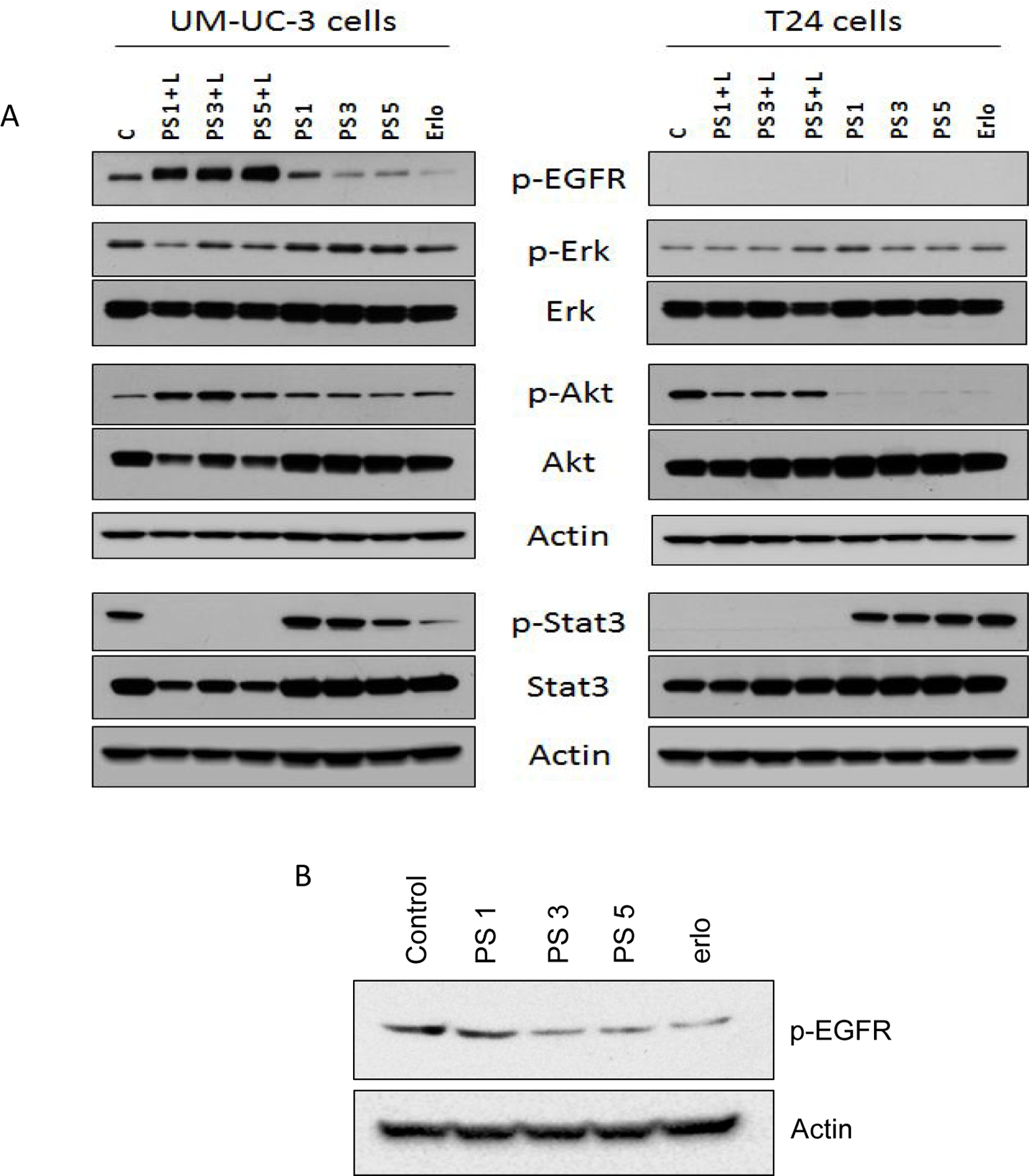
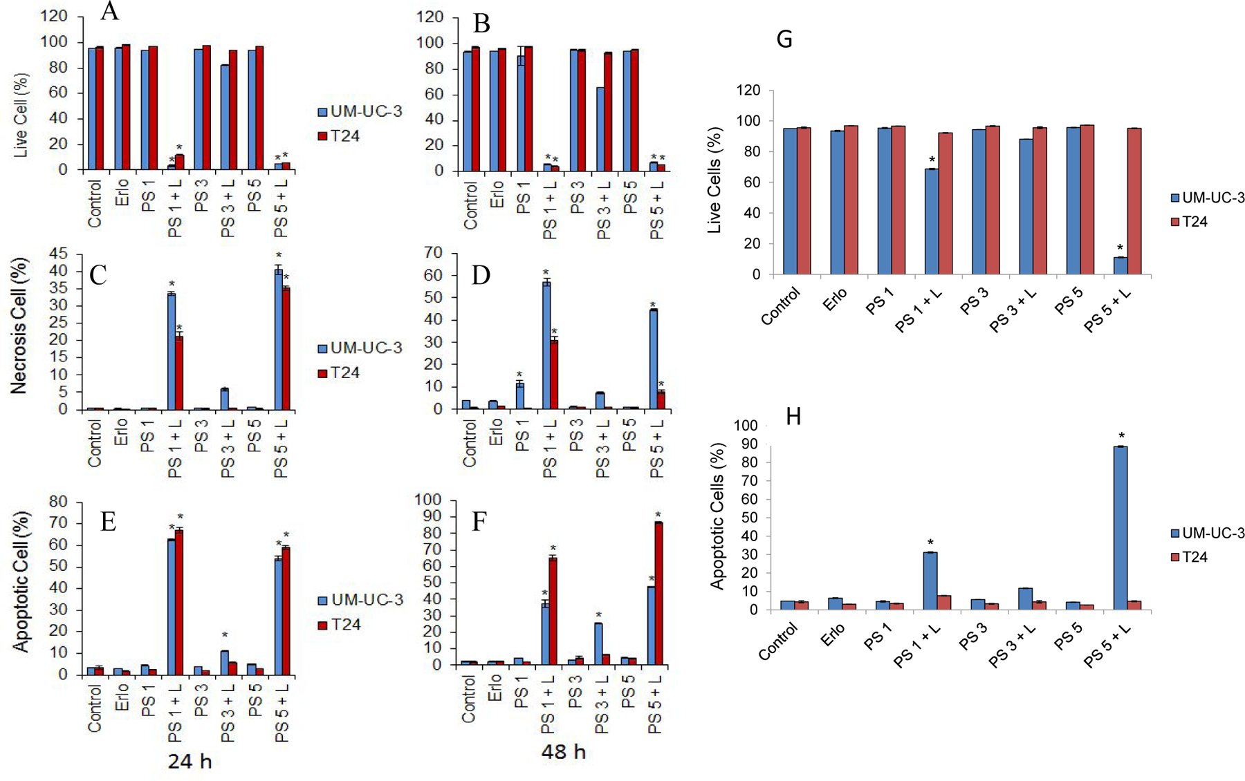
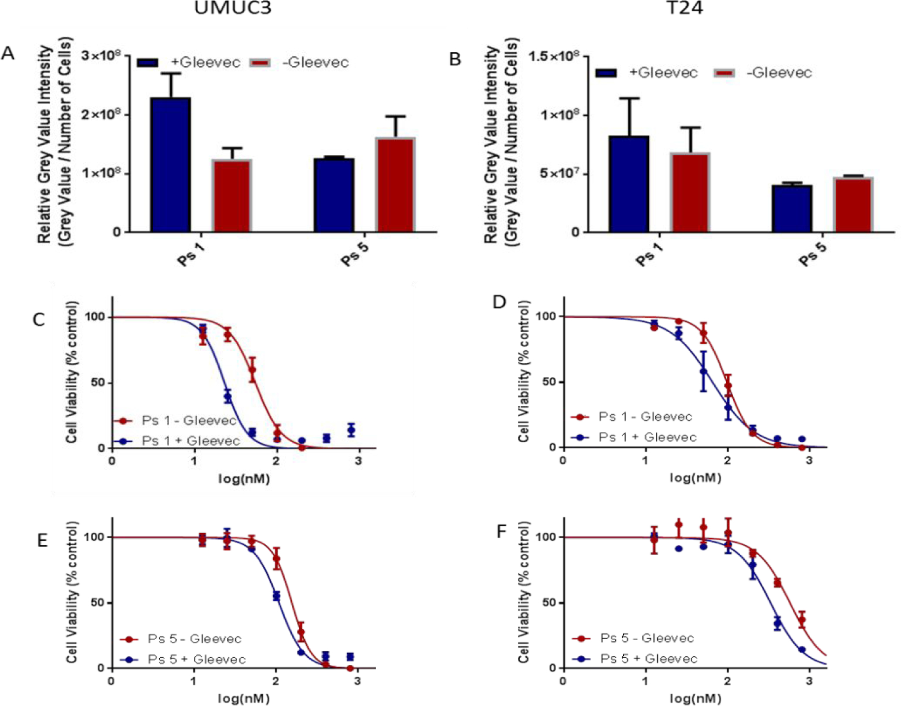
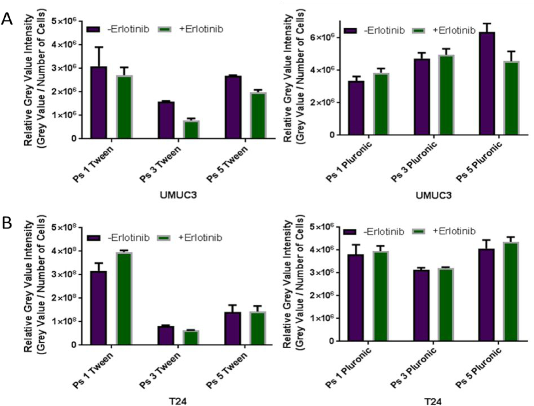
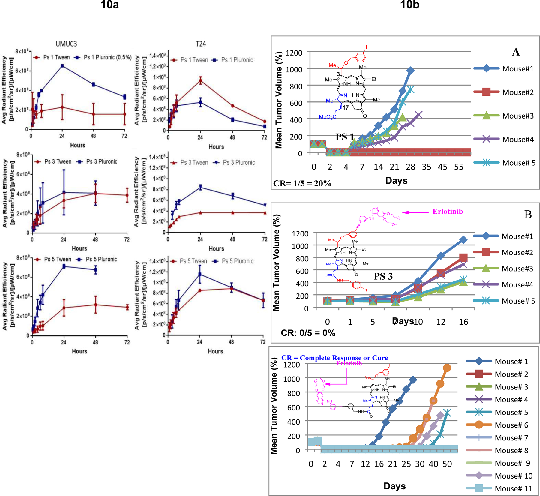
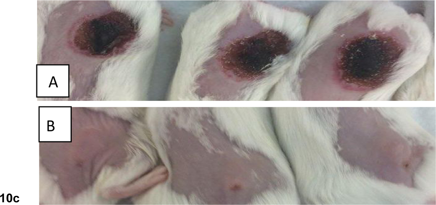
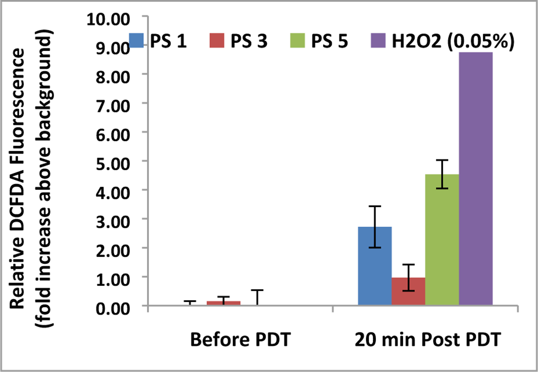
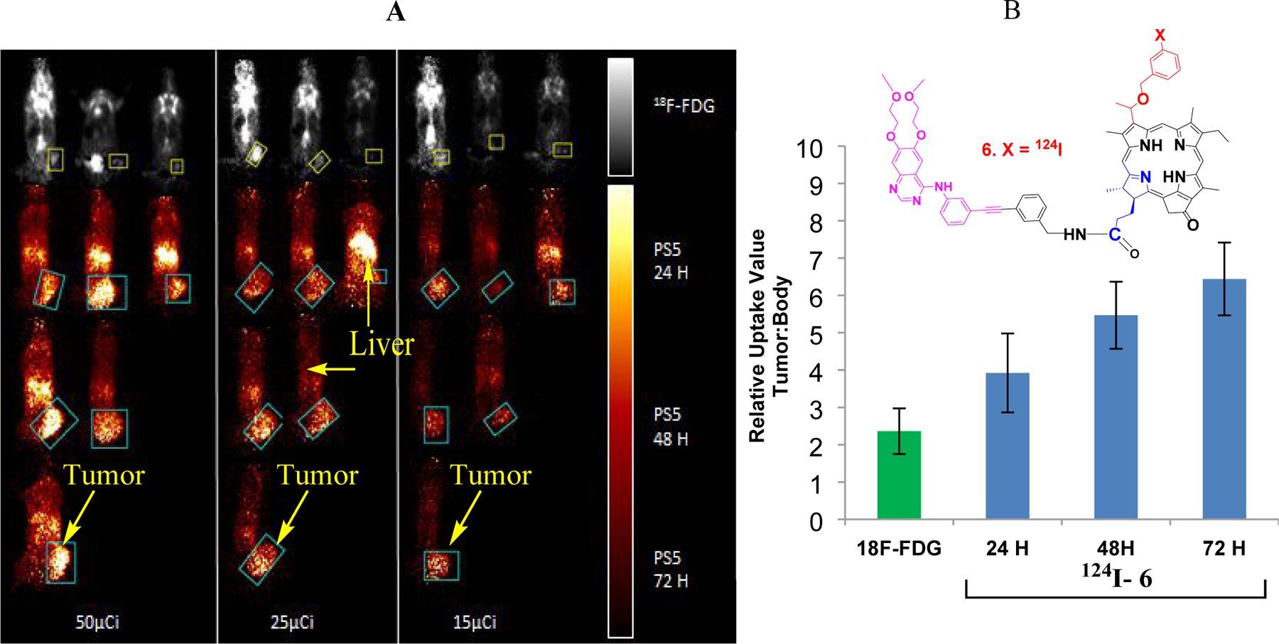
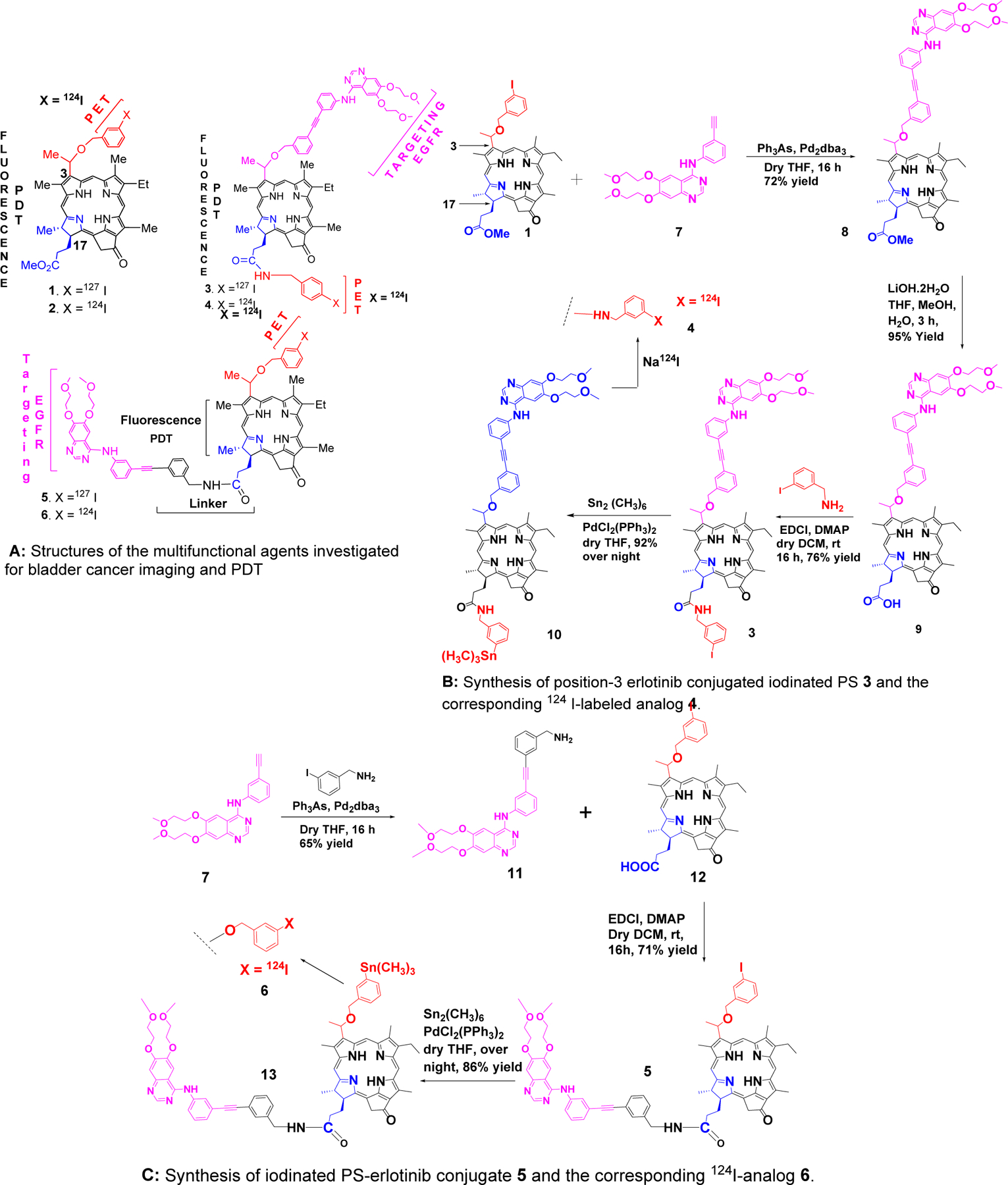
The in vitro and in vivo anticancer activity of iodinated photosensitizers (PSs) with and without an erlotinib moiety was investigated in UMUC3 [epidermal growth factor (EGFR)-positive] and T24 (EGFR-low) cell lines and tumored mice. Both the erlotinib-conjugated PSs 3 and 5 showed EGFR target specificity, but the position-3 erlotinib-PS conjugate 3 demonstrated lower photodynamic therapy efficacy than the corresponding non-erlotinib analogue 1, whereas the conjugate 5 containing an erlotinib moiety at position-17 of the PS showed higher tumor uptake and long-term tumor cure (severe combined immunodeficient mice bearing UMUC3 tumors). PS-erlotinib conjugates in the absence of light were ineffective in vitro and in vivo, but robust apoptotic and necrotic cell death was observed in bladder cancer cells after exposing them to a laser light at 665 nm. In contrast to 18F-fluorodeoxyglucose, a positron emission tomography agent, the position-17 erlotinib conjugate (124I-analogue 6) showed enhanced UMUC3 tumor contrast even at a low imaging dose of 15 μCi/mouse.


Figures

Figure 1
Figure 1
A: 1H NMR spectral regions of 1, 3 and 5 show significant chemical shift differences for specific protons. In 5, protons in the vicinity of the photosensitizer moiety’s C-ring (10-H, 132-CHH, and 12-CH3) are significantly shielded with respect to the corresponding protons of 1 and 3. Also, 5-H of the erlotinib moiety of 3 exhibits a substantial shielding compared to the 5-H chemical shift observed in 5. These observations suggest structural differences that are dependent on the erlotinib orientation in the conjugates (See text for details). B: 1H NMR spectra of 5 at 28 °C (bottom) and 55 °C (top). At elevated temperature, the chemical shifts of the 10-H (open triangle), 131-CHH (filled triangle), and 12-CH3 (arrow) proton signals exhibit a marked deshielding towards the positions observed for the corresponding protons of 1 and 3. This behavior is consistent with the breaking of pi-pi stacking interactions that may be responsible for the unusual shieldings observed for these protons in 5. (See figure in supporting information for additional spectra taken at other temperatures.)
Figure 2:
Figure 2:
Photophysical properties of photosensitizers with and without erlotinib moiety
Figure 3:
Figure 3:
Impact of solvents in absorption and fluorescence spectra of photosensitizers with and without erlotinib moiety.
Figure 4:
Figure 4:
Comparative in vitro photosensitizing efficacy of the photosensitizers formulated in Tween80 and Pluronic formulations.
Figure 5:
Figure 5:
Comparative uptake and intracellular localization (mitochondria vs. lysosomes) of PSs 1, 3, 5 formulated either in Tween80 or Pluronic127F in UMUC3 and T24 cells. The uptake PSs in Tween (A) and Pluronic (B) after 24 h incubation, and the fluorescence was measured using a CARY fluorimeter. The degree of fluorescence in each cell was directly compared as a measure of uptake. The fluorescence of each cell was measured using an Image Stream. Simultaneously, cells were stained with lysosphere green and mitotracker red to determine the subcellular localization specificity the PSs: (C) PSs 1, 3 & 5 Tween80 formulation in UMUC3, (D) PS 1, 3 & 5 Tween formulation in T24, (E) PSs 1, 3 & 5 Pluronic formulation in UMUC3 and (F) PS 1, 3 & 5 Pluronic formulation in T 24 cell line. The higher bright detail similarity score to a particular site reflects stronger overlap of the PS fluorescence to that site, either lysosomes or the mitochondria.
Figure 6:
Figure 6:
Comparative EGFR signal inhibition efficacy of erlotinib, PS 1 and the corresponding erlotinib conjugates 3 & 5 in bladder cancer cell lines UMUC-3 and T24 cell lines known for high and low expression of EGFR. (A) PS-erlotinib conjugates were treated at 1 µM concentration for 24 hours followed by PDT. (B) UM-UC-3 cells were treated with PS-erlotinib conjugates at 100 nM concentration for 24 hours.
Figure 7:
Figure 7:
Erlotinib conjugates induce robust apoptotic and necrotic cell death upon PDT. UMUC-3 and T24 cells were treated with different erlotinib conjugates (1.0 μM) with and without PDT. Cell death was quantified using trypan blue assay (A and B). Necrotic (C and D) and Apoptotic (E and F) cell death was quantified using Annexin V/PI labeling. Live cells and Apoptotic cell death was quantified in UM-UC-3 and T24 cells treated with erlotinib conjugates (100 nM) with and without PDT (G and H).
Figure 8:
Figure 8:
ABCG2 substrate specificity of PS with and without Erlotinib in UMUC3 and T24 cells. (A & B)) the uptake of PS 1 and 5 with and without the addition of Gleevec (small molecule inhibitor known to inhibit ABCG2 function) in UMUC3 and T24 cells. Cells were incubated with 500 nm of the Photosensitizers with or without the addition of Gleevec at 1uM concentration. (C-F) The effect of Gleevec on the toxicity of PS 1 and PS 5 at 48 h post incubation (light dose: I J).
Figure 9:
Figure 9:
Erlotinib competition with PSs 1, 3, and 5 in UMUC3 and T24 cells. UMUC3 (A) or T24 (B) cells were incubated with photosensitizer at 1 uM or Erlotinib at 3 uM for 4 hours. The cells were washed with warmed PBS three times then imaged using a Zeis Fluorescence microscope.
Figure 10
Figure 10
a: Comparative uptake of PS 1, 3, 5 (formulated either in Tween or Pluronic) in tumor, liver and skin (SCID mice bearing UMUC3 and T24 tumors) at a dose of 0.47 μmol/kg)) at variable time points (λex: 675 nm, λem: 720), using the IVIS optical imaging system. b: Comparative in vivo PDT efficacy (long-term antitumor activity) of PS with and without erlotinib conjugates. (A): PS 1 (non-erlotinib PS), (B): PS 3 erlotinib moiety is attached at position-3 of the PS (C): PS 5, erlotinib moiety is attached at position-17 of the PS. PS 1, 3, and 5 were individually injected (dose: 0.47 micromole/Kg) to SCID mice bearing UMUC-3 tumors at the flank. Mice were exposed to light (665 nm, 135 J/cm2, 75 mW/cm2) at 24 h post-injection (optimal uptake time) and tumor re-growth was monitored daily. These results suggest that position of the erlotinib moiety in the PS makes a remarkable difference in long-term tumor cure. c: Comparative tumor necrosis of mice injected with (A) PS 3 (effective PDT agent), and (B) PS 5 that showed limited efficacy at 72h post-light exposure. Mice were treated under similar drug and light doses. Dug dose: 0.47 μmol/kg, light dose: 135 J/cm2, 75 mW/cm2 and the tumors were exposed to light (665 nm) at 24h post-injection of the PS.
Figure 10
Figure 10
a: Comparative uptake of PS 1, 3, 5 (formulated either in Tween or Pluronic) in tumor, liver and skin (SCID mice bearing UMUC3 and T24 tumors) at a dose of 0.47 μmol/kg)) at variable time points (λex: 675 nm, λem: 720), using the IVIS optical imaging system. b: Comparative in vivo PDT efficacy (long-term antitumor activity) of PS with and without erlotinib conjugates. (A): PS 1 (non-erlotinib PS), (B): PS 3 erlotinib moiety is attached at position-3 of the PS (C): PS 5, erlotinib moiety is attached at position-17 of the PS. PS 1, 3, and 5 were individually injected (dose: 0.47 micromole/Kg) to SCID mice bearing UMUC-3 tumors at the flank. Mice were exposed to light (665 nm, 135 J/cm2, 75 mW/cm2) at 24 h post-injection (optimal uptake time) and tumor re-growth was monitored daily. These results suggest that position of the erlotinib moiety in the PS makes a remarkable difference in long-term tumor cure. c: Comparative tumor necrosis of mice injected with (A) PS 3 (effective PDT agent), and (B) PS 5 that showed limited efficacy at 72h post-light exposure. Mice were treated under similar drug and light doses. Dug dose: 0.47 μmol/kg, light dose: 135 J/cm2, 75 mW/cm2 and the tumors were exposed to light (665 nm) at 24h post-injection of the PS.
Figure 11:
Figure 11:
Comparative DCFDA fluorescence before and after exposure to 665 nm light. Plates were exposed to a total of 1 J of light (665 nm) at a rate of 3.2 mW/cm2.. Fluorescence value of cells with no DCFDA (background) was subtracted from before and after PDT. 0.05% H2O2 (30% stock was diluted to 0.18%, then 20 µL was added to each well) was added to 4 wells as a control before the plates were exposed to light. Photosensitizers were formulated in Tween80 formulation.
Figure 12.
Figure 12.
(A): PET images of SCID mice bearing UMUC3 tumors using 124I-6 and 18F-FDG. The images were analyzed using Amide Medical Imaging Software Boxes indicate Volume of Interest (VOI) in the coronal plane. Separate scale bars for 18F-FDG and 124 I-6 images are indicated on the right. The maximum threshold for color distribution was set to 10% in the 18F-FDG images and 50% in the 124I- 6 images. In each panel, the top row of mice show 18F-FDG uptake in the bladder, brain, chest and muscles, while tumor uptake is difficult to visualize. Tumors are well-visualized by 124I- 6 at alltime points, and uptake is primarily in the tumor and liver. 124I- 6 is retained in the tumor longer than in other parts of the body. (B): Relative Uptake Values were calculated as maximum tumor uptake divided by average whole body uptake. 18F- FDG (left) and 124I- 6 (right) RUVs indicate almost twice the specific uptake of 124I- 6 at 24 hours as compared to 18F-FDG. For II124 I- 6, the RUVs continue to rise at longer time points as it retains well in the tumor and less in the rest of the body.
Scheme 1:
Scheme 1:
Structures of parent compounds (radioactive and non-radioactive) derived from chlorophyll-a, and the synthesis of the corresponding erlotinib conjugates.

References

    1. Siegel R; Ma J; Zou Z; Jemal A Cancer Statistics, 2014. CA Cancer J Clin 2014, 64, 9–29. - PubMed
    1. Kaufman DS; Shipley WU; Feldman AS Bladder Cancer. Lancet 2009, 374, 239–249. - PubMed
    1. American Cancer Society: Bladder Cancer; Survival rates for bladder cancer by stage, 2016. Available at http://www.cancer.org/cancer/bladdercancer/detailedguide/bladder-cancer-... (accessed December 10, 2014).
    1. Grossman HB; Natale RB; Tangen CM; Speights VO; Vogelzang NJ; Trump DL; deVere White RW; Sarosdy MF; Wood DP; Raghavan D and Crawford ED, Neoadjuvant chemotherapy plus cystectomy compared with cystectomy alone for locally advanced bladder cancer. N Engl. J. Med 2003, 349, 859–866 - PubMed
    1. Hinata N; Hussein AA; George S; Trump DL; Levine EG; Omar K; Dasgupta P; Khan MS; Hosseini A; Wiklund P; Guru KA Impact of suboptimal neoadjuvant chemotherapy on peri-operative outcomes and survival after robot-assisted radical cystectomy: a multicentre multinational study. BJU Int 2017, 119(4), 605–611. - PubMed

Publication types

MeSH terms