. 2019 Apr:129:69-78.
doi: 10.1016/j.yjmcc.2019.02.009. Epub 2019 Feb 15.

Cardiac-specific deletion of GCN5L1 restricts recovery from ischemia-reperfusion injury

Affiliations

Cardiac-specific deletion of GCN5L1 restricts recovery from ischemia-reperfusion injury

Janet R Manning et al. J Mol Cell Cardiol. 2019 Apr.

Abstract

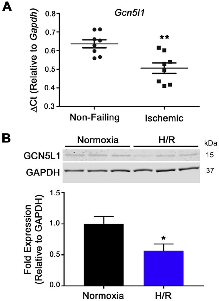
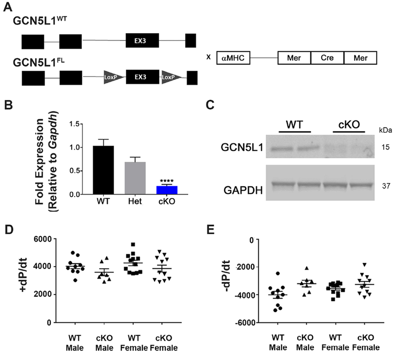
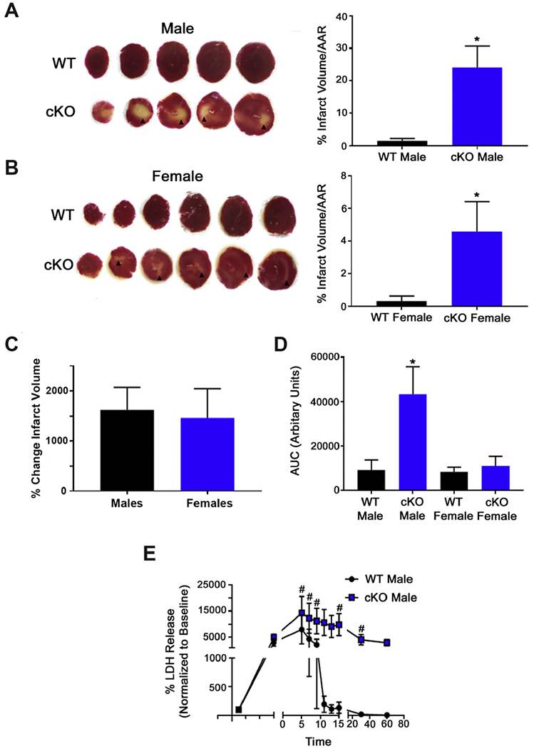
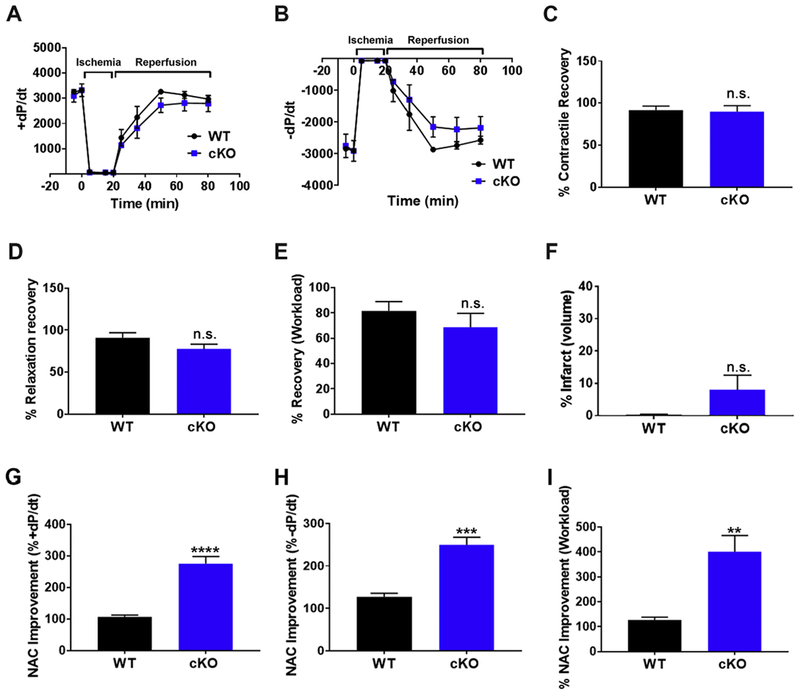
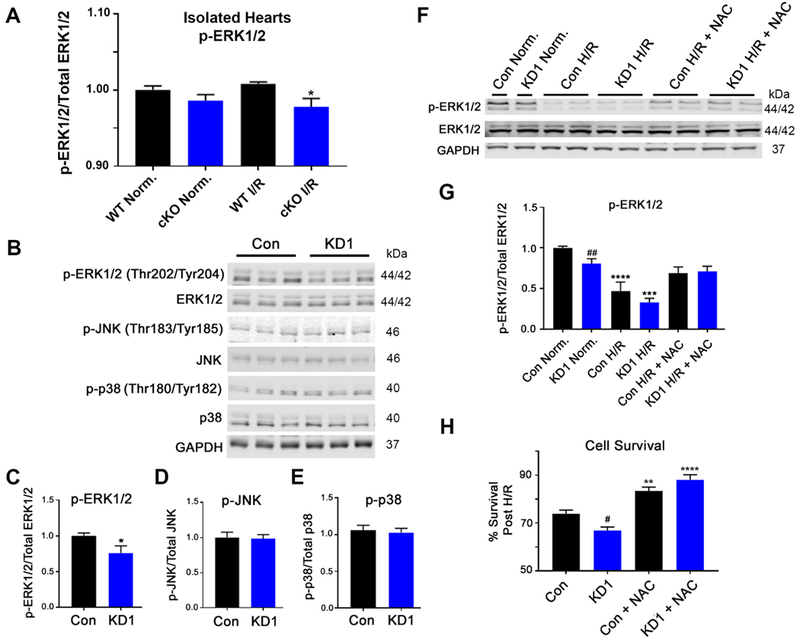
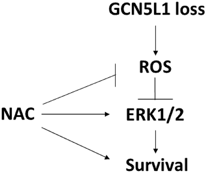
GCN5L1 regulates mitochondrial protein acetylation, cellular bioenergetics, reactive oxygen species (ROS) generation, and organelle positioning in a number of diverse cell types. However, the functional role of GCN5L1 in the heart is currently unknown. As many of the factors regulated by GCN5L1 play a major role in ischemia-reperfusion (I/R) injury, we sought to determine if GCN5L1 is an important nexus in the response to cardiac ischemic stress. Deletion of GCN5L1 in cardiomyocytes resulted in impaired myocardial post-ischemic function and increased infarct development in isolated work-performing hearts. GCN5L1 knockout hearts displayed hallmarks of ROS damage, and scavenging of ROS restored cardiac function and reduced infarct volume in vivo. GCN5L1 knockdown in cardiac-derived AC16 cells was associated with reduced activation of the pro-survival MAP kinase ERK1/2, which was also reversed by ROS scavenging, leading to restored cell viability. We therefore conclude that GCN5L1 activity provides an important protection against I/R induced, ROS-mediated damage in the ischemic heart.

Keywords: ERK1/2; Ex vivo working heart; GCN5L1; Ischemia reperfusion; Reactive oxygen species.


Conflict of interest statement

CONFLICTS OF INTEREST

None to report.

Figures

Figure 1.
Figure 1.. Ischemia downregulates GCN5L1 expression.
(A) GCN5L1 expression in human heart tissue from non-failing and chronic ischemic failing hearts normalized to Gapdh. N = 8 per group. (B) Immunoblot and quantitation of GCN5L1 protein from a human cardiomyocyte-derived cell line subjected to acute hypoxia and reoxygenation. N=6. * = P < 0.05, ** = P < 0.01, vs. control.
Figure 2.
Figure 2.. Development of cardiac-specific GCN5L1 knockout (cKO) mice.
(A) Mice with LoxP sites surrounding exon 3 of GCN5L1 were crossed to αMHC-MerCreMer mice to produce tamoxifen-inducible, cardiomyocyte-specific knockouts of GCN5L1 (GCN5L1 cKO). (B, C) MerCreMer expression in GCN5L1FL/FL cardiomyocytes leads to significant reductions in GCN5L1 transcript and protein expression. (D, E) No overt contractile phenotype is observed in the isolated hearts of cardiac-specific inducible knockouts. N= 5-12. **** = P < 0.0001 vs. control.
Figure 3.
Figure 3.. Loss of GCN5L1 expression reduces functional recovery after ischemia-reperfusion (I/R) injury.
(A) Experimental I/R protocol. (B, C) Work performed by isolated hearts after I/R is significantly reduced in cKO hearts from males and females. (D, E) After 60 minutes reperfusion, workload and contractility (+dP/dt) are significantly reduced in both cKO males and females. (F) Relaxation rate (−dP/dt) is significantly decreased in cKO hearts after reperfusion in females and trends towards a decrease in males. N = 4-6 mice per group. * = P < 0.05, ** = P < 0.01, *** = P < 0.001, **** = P < 0.0001 vs. control.
Figure 4.
Figure 4.. GCN5L1 loss promotes infarct development and LDH release from the myocardium.
(A, B) Representative TTC-stained hearts from male and female mice. Viable tissue is stained red. Arrowheads indicate infarcted myocardium. (C) Comparison of the change in infarct size caused by GCN5L1 loss in males and females. (D) Total cumulative LDH release (area under the curve) in male and female mice. (E) LDH release from male mice over the course of reperfusion. N = 4-6 mice per group. * = P < 0.05 vs. WT; # = P < 0.05 vs. baseline
Figure 5.
Figure 5.. Oxidative damage is increased in GCN5L1 cKO hearts.
(A) Protein carbonylation is elevated in both sham and I/R-subjected GCN5L1 cKO hearts. ** = P < 0.01 vs. wildtype. N = 5 mice per group. (B) Immunoblot confirmation of GCN5L1 knockdown (KD) in AC16 human ventricular myocytes using stably-expressed shRNA. N = 6. (C) Mitochondrial ROS production in control and GCN5L1 KD cells in normoxic and H/R conditions. (D-E) Seahorse XF analyses reveal differences in uncoupled oxygen consumption rates (OCR) and spare respiratory capacity. N = 36-41 replicates. (F) Incubation with mitoTEMPOL restores cell survival of KD cells in response to H/R injury. N = 6 per condition. ** = P < 0.01, *** = P < 0.001, **** = P < 0.0001 vs. normoxia, # = P < 0.05 vs. control.
Figure 6.
Figure 6.. Reducing ROS damage in GCN5L1 cKO hearts restores post-ischemic functional recovery.
(A, B) +dP/dt and −dP/dt recovery from I/R injury is restored in GCN5L1 cKO mice in the presence of ROS-scavenger N-acetyl cysteine (NAC). (C-E) Recovery after reperfusion in the presence of NACasa percentage of +dP/dt, −dP/dt, and workload. (F) Infarct volume is reduced three-fold in cKO mice in the presence of NAC following I/R injury, and is no longer significantly different from wildtype mice. (G-I) Changes in post-ischemic function relative to untreated hearts from Figure 3. N = 5 mice per group. ** = P < 0.01, *** = P < 0.001, **** = P <0.0001 vs. WT.
Figure 7.
Figure 7.. ROS scavenging restores pro-survival ERK1/2 signaling and cell viability in cardiac-derived GCN5L1 knockdown (KD) cells.
(A) ERK1/2 signaling is reduced in GCN5L1 cKO hearts following I/R injury. (B-E) Basal ERK1/2 signaling is reduced in GCN5L1 KD cells, while there is no change in the other MAP kinase proteins JNK or p38. N = 8-12 replicates per group. * = P < 0.05 vs. control. (F, G) ERK1/2 phosphorylation is significantly blunted in GCN5L1 KD cells at baseline, and in control and KD cells in response to H/R. Treatment with NAC restored ERK1/2 signaling in KD cells to baseline levels. (H) As with the isolated cKO hearts, treatment with NAC restored cell via bility in GCN5L1 KD cells subjected to H/R. N = 8-12 replicates per group. ** = P < 0.01, *** = P < 0.001, **** = P < 0.0001 vs. normoxia, # = P < 0.05, ## = P < 0.01 vs. control.
Figure 8.
Figure 8.. Hypothesized mechanism of action by GCN5L1 in cardiac cells following I/R injury.
Loss of GCN5L1 disrupts mitochondrial function leading to increased ROS production. Treatment of cells with antioxidants rescues GCN5L1-deficient cells after ischemic injury.

References

    1. Benjamin EJ et al. Heart Disease and Stroke Statistics—2017 Update: A Report From the American Heart Association. Circulation 135, (2017). - PMC - PubMed
    1. Brookes PS, Yoon Y, Robotham JL, Anders MW & Sheu S-S Calcium, ATP, and ROS: a mitochondrial love-hate triangle. Am. J. Physiol. - Cell Physiol 287, C817–C833 (2004). - PubMed
    1. Garcia-Rivas GJ & Torre-Amione G Abnormal mitochondrial function during ischemia reperfusion provides targets for pharmacological therapy. Methodist Debakey Cardiovasc. J 5, 2–7 (2009). - PubMed
    1. Shintani-Ishida K, Inui M & Yoshida K-I Ischemia-reperfusion induces myocardial infarction through mitochondrial Ca2+ overload. J. Mol. Cell. Cardiol 53, 233–239 (2012). - PubMed
    1. Alam MR, Baetz D & Ovize M Cyclophilin D and myocardial ischemia–reperfusion injury: A fresh perspective. J. Mol. Cell. Cardiol 78, 80–89 (2015). - PubMed

Publication types

MeSH terms