

Corneal Higher-Order Aberrations in Descemet Membrane Endothelial Keratoplasty versus Ultrathin DSAEK in the Descemet Endothelial Thickness Comparison Trial: A Randomized Clinical Trial
- PMID: 30776384
- PMCID: PMC6590707
- DOI: 10.1016/j.ophtha.2019.02.007
Corneal Higher-Order Aberrations in Descemet Membrane Endothelial Keratoplasty versus Ultrathin DSAEK in the Descemet Endothelial Thickness Comparison Trial: A Randomized Clinical Trial
Abstract
Purpose: To compare corneal higher-order aberrations (HOA) after ultrathin Descemet stripping automated endothelial keratoplasty (DSAEK) and Descemet membrane endothelial keratoplasty (DMEK).
Design: Patient- and outcome-masked randomized controlled clinical trial.
Participants: Patients with damaged or diseased endothelium from Fuchs endothelial dystrophy or pseudophakic bullous keratopathy who were good candidates for DMEK or ultrathin DSAEK.
Methods: Corneal anterior and posterior surface HOA were measured with Scheimpflug imaging before surgery and at 3, 6, and 12 months after surgery. HOA after ultrathin DSAEK and DMEK were compared; correlation was performed between best spectacle-corrected visual acuity (BSCVA) and HOA at each time point.
Main outcome measures: Higher-order aberrations of the anterior and posterior cornea, expressed as the root mean square deviation from a best fit sphere reference surface.
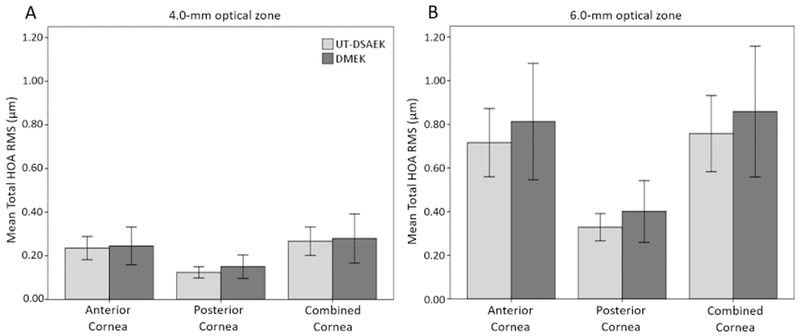
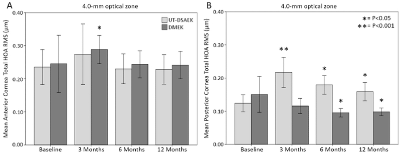
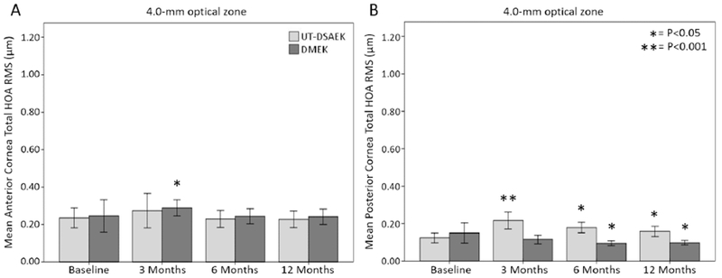
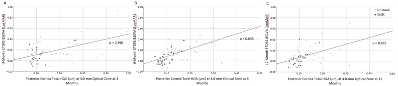
Results: At 3, 6, and 12 months after surgery, the posterior corneal surface had significantly less coma (P ≤ 0.003) and total HOA (P ≤ 0.001) in DMEK compared with ultrathin DSAEK (4.0- and 6.0-mm OZ). Posterior trefoil (P ≤ 0.034), secondary astigmatism (P ≤ 0.042), and tetrafoil (P ≤ 0.045) were lower in DMEK than ultrathin DSAEK at 3, 6, or 12 months (either 4.0- or 6.0-mm OZ). There were no significant differences in anterior surface HOA between DMEK and ultrathin DSAEK at any post-surgical time. Compared with baseline, total posterior HOA was increased (P ≤ 0.036) in ultrathin DSAEK at 3, 6, and 12 months, in contrast to DMEK, where it was decreased (P ≤ 0.044) at 6 and 12 months (4.0- or 6.0-mm OZ, or both). At 6 and 12 months, posterior corneal total HOA correlated with BSCVA (ρ ≤ 0.635, P ≤ 0.001; 4.0- and 6.0-mm OZ). There were no moderate or strong correlations between anterior or combined corneal surface HOA at any time point after surgery.
Conclusions: Descemet membrane endothelial keratoplasty results in less posterior corneal HOA compared with ultrathin DSAEK. Descemet membrane endothelial keratoplasty decreases and ultrathin DSAEK increases posterior corneal HOA compared with presurgical values. Total posterior corneal HOA correlates with 6- and 12-month postoperative visual acuity and may account for the better visual acuity observed after DMEK.
Copyright © 2019 American Academy of Ophthalmology. Published by Elsevier Inc. All rights reserved.
Conflict of interest statement
Figures
Similar articles
-
Ultrathin-Descemet Stripping Automated Endothelial Keratoplasty Versus Descemet Membrane Endothelial Keratoplasty: A Systematic Review and Meta-analysis.In Vivo. 2023 Jan-Feb;37(1):400-409. doi: 10.21873/invivo.13092. In Vivo. 2023. PMID: 36593036 Free PMC article.
-
Descemet Endothelial Thickness Comparison Trial: A Randomized Trial Comparing Ultrathin Descemet Stripping Automated Endothelial Keratoplasty with Descemet Membrane Endothelial Keratoplasty.Ophthalmology. 2019 Jan;126(1):19-26. doi: 10.1016/j.ophtha.2018.05.019. Epub 2018 Jun 23. Ophthalmology. 2019. PMID: 29945801 Clinical Trial.
-
Mediators of Visual Acuity in Descemet Membrane Endothelial Keratoplasty and Ultrathin Descemet Stripping Automated Endothelial Keratoplasty.Cornea. 2024 Jul 1;43(7):853-859. doi: 10.1097/ICO.0000000000003434. Epub 2023 Nov 21. Cornea. 2024. PMID: 37986182 Clinical Trial.
-
Descemet Membrane Endothelial Keratoplasty versus Ultrathin Descemet Stripping Automated Endothelial Keratoplasty: A Multicenter Randomized Controlled Clinical Trial.Ophthalmology. 2020 Sep;127(9):1152-1159. doi: 10.1016/j.ophtha.2020.02.029. Epub 2020 Mar 2. Ophthalmology. 2020. PMID: 32386811 Clinical Trial.
-
Ultrathin Descemet Stripping Automated Endothelial Keratoplasty (UT-DSAEK) versus Descemet Membrane Endothelial Keratoplasty (DMEK)-a systematic review and meta-analysis.Eye (Lond). 2023 Oct;37(14):3026-3032. doi: 10.1038/s41433-023-02467-2. Epub 2023 Mar 18. Eye (Lond). 2023. PMID: 36934158 Free PMC article.
Cited by
-
Screening and Grading of Textural Interface Opacities in DSAEK Grafts with the M-TIO Scale for Predicting Visual Outcomes.Diagnostics (Basel). 2025 May 14;15(10):1241. doi: 10.3390/diagnostics15101241. Diagnostics (Basel). 2025. PMID: 40428234 Free PMC article.
-
Higher Order Aberrations following Scleral Buckling Surgery in Patients with Rhegmatogenous Retinal Detachment.Healthcare (Basel). 2021 Nov 27;9(12):1643. doi: 10.3390/healthcare9121643. Healthcare (Basel). 2021. PMID: 34946371 Free PMC article.
-
Collagen Remodeling Plays a Pivotal Role in Endothelial Corneal Dystrophies.Invest Ophthalmol Vis Sci. 2020 Dec 1;61(14):1. doi: 10.1167/iovs.61.14.1. Invest Ophthalmol Vis Sci. 2020. PMID: 33259606 Free PMC article.
-
Ultrathin-Descemet Stripping Automated Endothelial Keratoplasty Versus Descemet Membrane Endothelial Keratoplasty: A Systematic Review and Meta-analysis.In Vivo. 2023 Jan-Feb;37(1):400-409. doi: 10.21873/invivo.13092. In Vivo. 2023. PMID: 36593036 Free PMC article.
-
Corneal Light Scatter After Ultrathin Descemet Stripping Automated Endothelial Keratoplasty Versus Descemet Membrane Endothelial Keratoplasty in Descemet Endothelial Thickness Comparison Trial: A Randomized Controlled Trial.Cornea. 2020 Jun;39(6):691-696. doi: 10.1097/ICO.0000000000002256. Cornea. 2020. PMID: 31939923 Free PMC article. Clinical Trial.
References
-
- Patel S, Baratz K, Hodge D, et al. The effect of corneal light scatter on vision after Descemet stripping with endothelial keratoplasty. Arch Ophthalmol 2009;12:153–160. - PubMed
-
- Van Dijk K, Droutsas K, Hou J, et al. Optical quality of the cornea after Descemet membrane endothelial keratoplasty. Am J Ophthalmol 2014;158:71–79. e1. - PubMed
-
- Nielsen E, Ivarsen A, Kristensen S, Hjortdal J. Fuchs’ endothelial corneal dystrophy: a controlled prospective study on visual recovery after endothelial keratoplasty. Acta Ophthalmol 2016;94:780–787. - PubMed
-
- Rudolph M, Laaser K, Bachmann BO, et al. Corneal Higher-Order Aberrations after Descemet’s Membrane Endothelial Keratoplasty. Ophthalmology 2012;119:528–535. - PubMed
Publication types
MeSH terms
Grants and funding
LinkOut - more resources
Full Text Sources
Medical
