. 2019 Apr:129:242-249.
doi: 10.1016/j.micpath.2019.02.019. Epub 2019 Feb 15.

House dust mite exposure attenuates influenza A infection in a mouse model of pulmonary allergic inflammation

Affiliations

House dust mite exposure attenuates influenza A infection in a mouse model of pulmonary allergic inflammation

Qiyao Hu et al. Microb Pathog. 2019 Apr.

Abstract

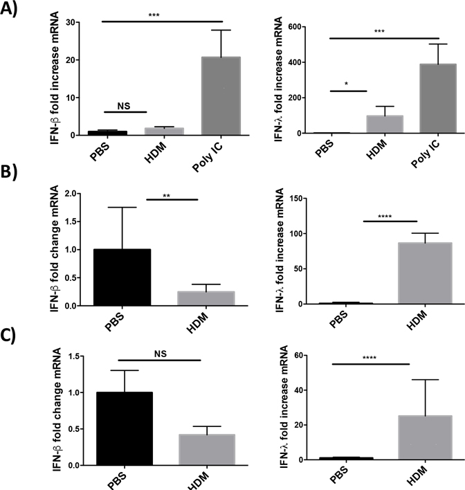
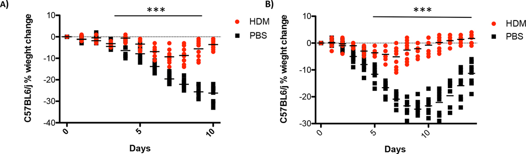
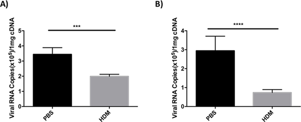
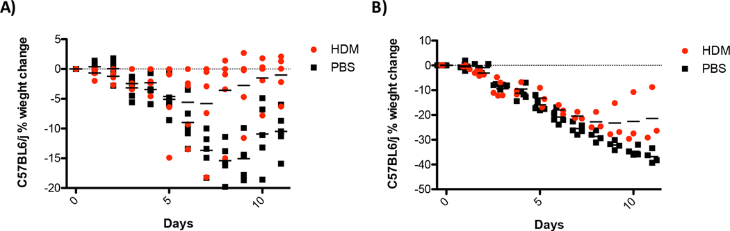
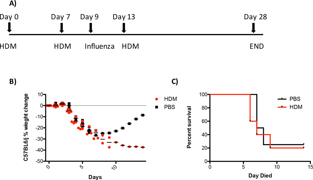
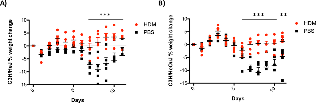
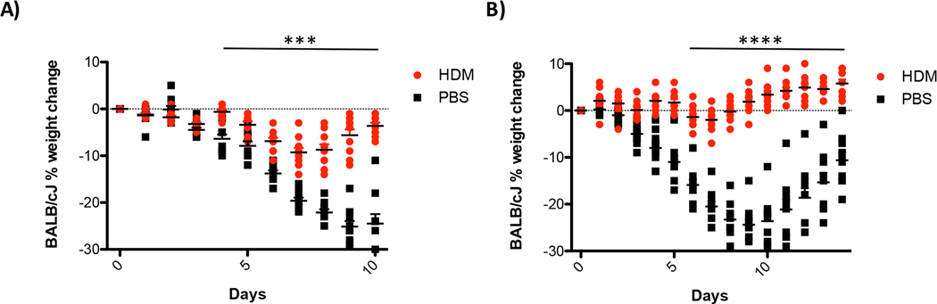
Environmental allergens elicit complex immune responses in the lungs that can promote the development of asthma or exacerbate preexisting asthma in susceptible individuals. House dust mites are one of the most common indoor allergens and are a significant driver of allergic disease. Respiratory infections are known factors in acute exacerbations of asthma but the impact of allergen on the pathogen is not well understood. We investigated the pathogenesis of influenza A infection following exposure to house dust mites. Mice exposed to house dust mites lose less weight following infection and had more transcription of interferon-lambda than controls. These data correlated with less transcription of the influenza polymerase acidic gene suggesting diminished viral replication in house dust mite exposed mice. Altogether, these data suggest that exposure to environmental allergens can influence the pathogenesis of influenza infection.

Keywords: Allergy; Asthma; House dust mite; Inflammation; Influenza.


Figures

Figure 1
Figure 1. House dust mite exposure scheme
A) short term exposure model, mice are exposed once to 100 μg of HDM IT and are infected with 400 PFU of influenza 18 hrs later. Control mice are treated with 100 μl saline IT and are then infected with influenza. B) long term exposure model, mice are exposed IT to 100 μg of HDM once a week for three weeks and are infected with 400 PFU of influenza virus 48hrs after the last HDM treatment. Control mice are exposed three times IT with 100 μl of saline and infected with 400 PFU 48hrs after the last exposure to saline.
Figure 2
Figure 2. House dust mite exposure induces interferon expression.
C57BL/6J mice were treated once with 100 μg of HDM or 100 μl of saline and lungs were harvested at 8hrs (A), 18hrs (B), or three times weekly (C) and lungs were harvested 48hrs after the last HDM treatment. Lungs were prepared for qRT-PCR and the expression of IFN-β and IFN-λ was determined using Taqman qRT-PCR. For the 8hr time point a positive control was included and mice were treated IT with 50ug of Poly I:C. Data is representative of two independent experiments with 3 mice per group. Data was analyzed by ANOVA with Dunnett’s multiple comparisons test **p=0.005, ***p=0.0005, NS not significant.
Figure 3
Figure 3. House dust mite exposure protects against influenza A infection.
C57BL/6J mice were treated once for 18hrs (A) or three times (B) with 100 μg of HDM or 100 μl of saline. Mice were challenged IT with 400 PFU of influenza and body weight was determined daily. Data represents 10 mice and was analyzed by ANOVA***p=0.005, ****p=0.0005.
Figure 4
Figure 4. Exposure to house dust mite reduces viral load.
C57BL/6J mice were exposed to HDM or saline IT once (A) or three times (B). Mice were infected IT with 400 PFU of influenza A and lungs were harvested 4 days after infection and prepared for Taqman qRT-PCR. As a marker for viral replication we determined the levels of polymerase acidic (PA) gene mRNA in the lungs of infected mice. Data is representative of two experiments N=3/group. Statistical significance was determined by ANOVA ***p=0.005, ****p=0.0005.
Figure 5
Figure 5. House dust mite exposure does not protect against a lethal influenza challenge.
C57BL/6J mice were exposed three times to 100 μg of house dust mites or saline as a control. Forty-eight hours after the last exposure to HDM or saline mice were infected with 4,000 PFU of influenza A. Body weight was monitored daily. N=5 mice/group, ANOVA determined statistical significance **** p=0.0005.
Figure 6
Figure 6. Chronic exposure to house dust mite protects against influenza infection.
Mice were exposed to HDM or saline weekly for eight weeks prior to infection with 400 or 4,000 PFU of influenza A. Body weights were monitored daily. N=5 mice/group ANOVA determined statistical significance ****p=0.0005. Note that statistical significance with a multiple measures ANOVA could not be determined in (B) due to unequal group sizes after day 5.
Figure 7
Figure 7. House dust mite exposure of sensitized animals after influenza infection worsens outcomes.
Mice were exposed to 100 μg of HDM or 100 μl of saline twice a week apart. Mice were then infected IT with 400 PFU of influenza virus 48hrs after the second exposure to HDM or saline. Four days after influenza virus infection mice were challenged with 100 μg of HDM or 100 μl of saline. Body weight was monitored daily. Mice were euthanized if body weight loss ≥ 30% of the initial body weight. (A) schematic of experiment (B) Body weight loss (C) data from (B) plotted as a survival graph. N=5 mice for the HDM group and N=4 for the saline group.
Figure 8
Figure 8. House dust mite-mediated protection from influenza infection is independent of TLR-4.
(A) TLR-4 deficient C3H/HeJ mice were exposed IT to 100 μg of HDM or 100 μl of saline weekly for three weeks. Forty-eight hours after the last exposure, mice were challenged with 400 PFU of influenza A. (B) TLR-4 sufficient C3H/HeOuJ were treated similarly. Body weight was monitored daily. N=5 mice/group ANOVA determined statistical significance **p=0.05, ****p=0.0005
Figure 9
Figure 9. HDM exposure protects BALB/cJ mice from influenza infection.
Mice prone to allergic inflammation, BALB/cJ, were evaluated for protection from influenza infection. Mice were treated as outlined in Figure 1. Both mice exposed once (A) or three times (B) were protected from influenza A virus infection. N= 10 mice/group statistical significance was determined by ANOVA ***p=0.005, ****p=0.0005.
Figure 10
Figure 10. Inflammatory histopathology of BALB/cJ mice infected with influenza
A. BALB/cJ mice were exposed to saline or HDM once or three times prior to being infected IT with 400 PFU of influenza A. Mice were randomized and 3 mice/treatment/time point were euthanized at the indicated time and lungs were removed for histological analysis. Inflammatory pathology was evaluated by investigators blinded to the treatment groups. Images of representative large airways were obtained at 10x magnification. Red arrows indicate inflammatory exudate in the conducting airways.

Similar articles

Cited by

References

    1. Akinbami LJ, Moorman JE, Bailey C, Zahran HS, King M, Johnson CA, et al. Trends in asthma prevalence, health care use, and mortality in the United States, 2001–2010. NCHS data brief. 2012;(94):1–8. . - PubMed
    1. Pawankar R Allergic diseases and asthma: a global public health concern and a call to action. World Allergy Organ J. 2014;7(1):12. doi: 10.1186/1939-4551-7-12. . - DOI - PMC - PubMed
    1. Ring J, Akdis C, Lauener R, Schappi G, Traidl-Hoffmann C, Akdis M, et al. Global Allergy Forum and Second Davos Declaration 2013 Allergy: Barriers to cure--challenges and actions to be taken. Allergy. 2014;69(8):978–82. doi: 10.1111/all.12406. - DOI - PubMed
    1. Hamid Q, Tulic M. Immunobiology of asthma. Annu Rev Physiol. 2009;71:489–507. Epub 2009/07/07. doi: 10.1146/annurev.physiol.010908.163200. - DOI - PubMed
    1. Galli SJ, Tsai M, Piliponsky AM. The development of allergic inflammation. Nature. 2008;454(7203):445–54. doi: 10.1038/nature07204. - DOI - PMC - PubMed