. 2019 Jul;144(1):183-192.
doi: 10.1016/j.jaci.2019.01.045. Epub 2019 Feb 16.

HLA-A*32:01 is strongly associated with vancomycin-induced drug reaction with eosinophilia and systemic symptoms

Affiliations

HLA-A*32:01 is strongly associated with vancomycin-induced drug reaction with eosinophilia and systemic symptoms

Katherine C Konvinse et al. J Allergy Clin Immunol. 2019 Jul.

Abstract

Background: Vancomycin is a prevalent cause of the severe hypersensitivity syndrome drug reaction with eosinophilia and systemic symptoms (DRESS), which leads to significant morbidity and mortality and commonly occurs in the setting of combination antibiotic therapy, affecting future treatment choices. Variations in HLA class I in particular have been associated with serious T cell-mediated adverse drug reactions, which has led to preventive screening strategies for some drugs.

Objective: We sought to determine whether variation in the HLA region is associated with vancomycin-induced DRESS.

Methods: Probable vancomycin-induced DRESS cases were matched 1:2 with tolerant control subjects based on sex, race, and age by using BioVU, Vanderbilt's deidentified electronic health record database. Associations between DRESS and carriage of HLA class I and II alleles were assessed by means of conditional logistic regression. An extended sample set from BioVU was used to conduct a time-to-event analysis of those exposed to vancomycin with and without the identified HLA risk allele.

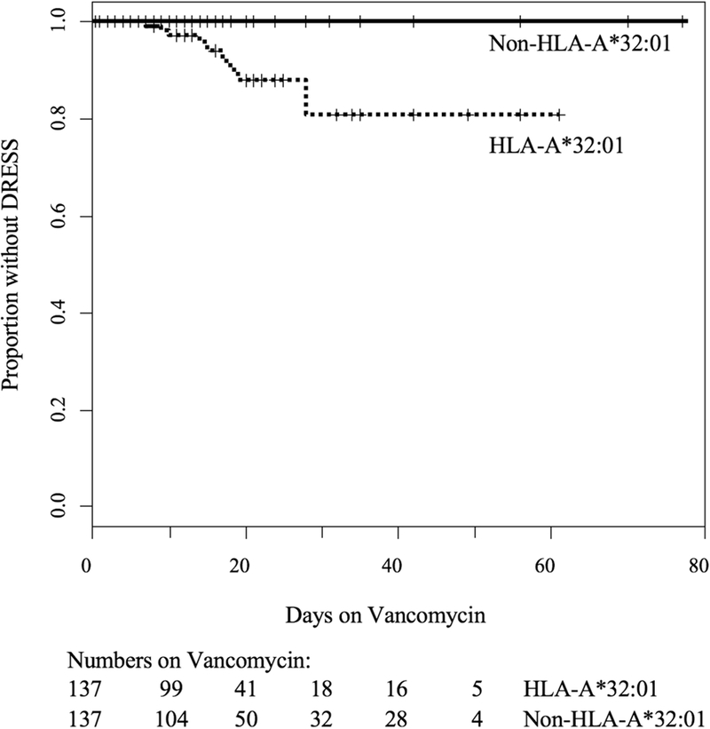
Results: Twenty-three subjects met the inclusion criteria for vancomycin-associated DRESS. Nineteen (82.6%) of 23 cases carried HLA-A*32:01 compared with 0 (0%) of 46 of the matched vancomycin-tolerant control subjects (P = 1 × 10⁻⁸) and 6.3% of the BioVU population (n = 54,249, P = 2 × 10-16). Time-to-event analysis of DRESS development during vancomycin treatment among the HLA-A*32:01-positive group indicated that 19.2% had DRESS and did so within 4 weeks.

Conclusions: HLA-A*32:01 is strongly associated with vancomycin-induced DRESS in a population of predominantly European ancestry. HLA-A*32:01 testing could improve antibiotic safety, help implicate vancomycin as the causal drug, and preserve future treatment options with coadministered antibiotics.

Keywords: T-cell hypersensitivity; Vancomycin; antibiotic allergy; delayed hypersensitivity; drug reaction with eosinophilia and systemic symptoms; human leukocyte antigen.


Conflict of interest statement

Conflicts of Interest: The authors have no conflicts of interest relevant to this manuscript. However, EP receives consulting fees from Biocryst. EP and SM receive royalties from UpToDate and have equity in IIID Pty Ltd that holds a patent for HLA-B*57:01 testing for abacavir hypersensitivity. MR serves as a consultant for Merck, Processa Pharma and aTyr Pharma and is a Deputy Editor for JAMA Dermatology.

Figures

Figure 1.
Figure 1.. HLA-A*32:01 is strongly associated with vancomycin DRESS.
A. 19/23 (83%) DRESS cases carried HLA-A*32:01 compared with 0/46 (0%) of the matched vancomycin tolerant controls (p=1×10⁻⁸, conditional logistic). If analyses are restricted to immunologically confirmed cases, then 11/12 (92%) vancomycin ELISpot positive patients carried HLA-A*32:01 compared with 0/24 (0%) of the BioVU matched controls (p=9×10⁻⁷, conditional logistic). HLA-A*32:01 carriage in all identified vancomycin DRESS cases and immunologically confirmed cases was also very significantly overrepresented compared to HLA-A*32:01 carriage in the entire BioVU cohort (6.3%) (p=2×10–16 and p=2=7×10–13 respectively, exact binomial tests). There was no significant difference in HLA-A*32:01 carriage between the vancomycin tolerant populations and the BioVU cohort (p=0.12 for all controls, p=0.40 for controls matched to immunologically confirmed cases, exact binomial tests). Additionally, there was no significant difference in HLA-A*32:01 carriage between the immunologically confirmed vancomycin DRESS cases and those that were not immunologically confirmed (p=0.32, Fisher’s exact test). All analyses shown included Bonferroni correction for multiple comparisons. B. IFN-γ release ELISpot results after 18-hour incubation with vancomycin at concentrations of 250 µg/mL (grey) or 500 µg/mL (black) using peripheral blood mononuclear cells from vancomycin DRESS patients. Controls included cells from vancomycin-naïve, HLA-A*32:01 positive healthy donors (n = 3) including the son of case patient 18 and the vancomycin skin test negative control C50, an HLA-A*32:01 positive individual tolerant of 4 weeks of vancomycin (n = 1), patients who had developed a non-DRESS immune-mediated adverse reaction to vancomycin (n = 5) and non-HLA matched healthy donors (n = 4). Means of the replicates are plotted. In patients with multiple blood draws at time points distant from the reaction, ELISpot results from the first blood draw are plotted. 12/14 (85.7%) DRESS cases had a positive vancomycin ELISpot compared to none of the controls (p=0.005 (DRESS vs. HLA-A-32:01 positive controls), p=0.002 (DRESS vs. non-DRESS ADRs), p=0.005 (DRESS vs. non-HLA matched healthy donors)). Positive results are those above the dotted line intersecting the y-axis at 50 SFU/million cells. Differences in proportion of positive responses between groups were assessed using Fisher’s exact tests. Patient and control PBMCs were also stimulated with vancomycin at concentrations of 5 µg/mL and 50 µg/mL and exhibited a dose-dependent response (data not shown). Legend: HLA, human leukocyte antigen; Vanc, vancomycin; DRESS, drug reaction with eosinophilia and systemic symptoms; SFU, spot-forming units; IM-ADR, immune-mediated adverse drug reaction.
Figure 2.
Figure 2.. Time-to-event analysis demonstrating that only HLA-A*32:01 positive patients developed DRESS.
Kaplan-Meier estimates were used to determine time to DRESS or possible DRESS development during vancomycin treatment stratified by carriage of HLA-A*32:01. Cases of DRESS occurred in HLA-A*32:01 positive subjects between 1 and 4 weeks of vancomycin therapy but not in HLA-A*32:01 negative subjects. The estimated risk of DRESS prior to 4 weeks of treatment was 19.2% in those carrying the HLA-A*32:01 allele.
Figure 3.
Figure 3.. Vancomycin skin testing, acute DRESS skin eruption and skin biopsy histology.
A and B. Vancomycin intradermal testing (IDT) results in patient 18 approximately 6.5 months after developing vancomycin DRESS and control C50, an HLA-A*32:01 positive, vancomycin-naïve healthy donor. IDT was performed on the volar forearm of the skin with 0.02 mL of vancomycin at concentrations of 0.05, 0.5, 5 and 50 mg/mL. The positive histamine and negative saline controls worked as expected. Vancomycin produced a strong immediate histamine response at 20 minutes in both control C50 and patient 18, but only patient 18 with a history of HLA-A*32:01 positive DRESS developed a concentration dependent induration of the skin at 48 hours at the 0.5, 5 and 50 mg/ml concentrations. Additionally, patient 18 had negative IDT to levofloxacin (not shown) and was successfully rechallenged with levofloxacin, a drug that, at the time of reaction, was administered with vancomycin. C. A representative example of the skin eruption from patient 18 during acute vancomycin DRESS. She had a diffuse morbilliform exanthema with facial involvement and facial edema (not shown). D. Hematoxylin and eosin staining of punch biopsies of skin from patient 18. Acute DRESS histology from a skin biopsy taken three days following onset of symptoms (upper panel) and skin test histology from the 5 mg/ml vancomycin positive intradermal skin test at 48 hours (lower panels) demonstrate papillary dermal edema, epidermal spongiosis and a dense lymphocytic infiltrate. Rare eosinophils are present on both biopsies. The skin test histology mirrors the results from the acute biopsy. E. Immunohistochemistry of T-cell subsets from acute DRESS and positive skin test biopsies from patient 18. Healthy skin is shown for comparison (upper panel). There was no discernable difference in the distribution of CD4 and CD8 positive cells in the dermal infiltrate between the acute (middle panel) and skin test (lower panel) biopsies. The number of intraepidermal CD8 positive cells was substantially higher in the acute biopsy. There was no appreciable exocytosis of CD4 T cells in the acute biopsy or CD4 or CD8 T cells in the biopsy from vancomycin IDT. Notably, dermal FOXP3+, regulatory T cells were present in the skin test biopsy, but absent in the acute biopsy.
Figure 4.
Figure 4.. Molecular docking prediction of vancomycin binding HLA-A*32:01.
Vancomycin is shown as sticks, white for carbon, blue for nitrogen, red for oxygen. Vancomycin atoms that mediate intermolecular contacts with D-Ala-D-Ala (in PDB 1FVM) are shown in cyan. A homology model of HLA-A*32:01 is shown in yellow as a ribbon diagram. Polymorphic positions that distinguish the associated HLA-A*32:01 allele from the closely related HLA-A*29:02 allele are shown in magenta. Intermolecular contacts between vancomycin and HLA-A*32:01 predicted by molecular docking are shown as black dashes.

Comment in

References

    1. Lin YF, Yang CH, Sindy H, Lin JY, Rosaline Hui CY, Tsai YC, et al. Severe cutaneous adverse reactions related to systemic antibiotics. Clin Infect Dis 2014;58(10):1377–85. - PubMed
    1. Blumenthal KG, Peter JG, Trubiano JA, Phillips EJ. Antibiotic allergy. Lancet 2018. - PMC - PubMed
    1. Pavlos R, Mallal S, Phillips E. HLA and pharmacogenetics of drug hypersensitivity. Pharmacogenomics 2012;13(11):1285–306. - PubMed
    1. Aota N, Shiohara T. Viral connection between drug rashes and autoimmune diseases: how autoimmune responses are generated after resolution of drug rashes. Autoimmun Rev 2009;8(6):488–94. - PubMed
    1. Ghislain PD, Roujeau JC. Treatment of severe drug reactions: Stevens-Johnson syndrome, toxic epidermal necrolysis and hypersensitivity syndrome. Dermatol Online J 2002;8(1):5. - PubMed

Publication types