

Segregation of salience network predicts treatment response of depression to repetitive transcranial magnetic stimulation
- PMID: 30776777
- PMCID: PMC6378906
- DOI: 10.1016/j.nicl.2019.101719
Segregation of salience network predicts treatment response of depression to repetitive transcranial magnetic stimulation
Abstract
Background: The present study tested the hypothesis that network segregation, a graph theoretic measure of functional organization of the brain, is correlated with treatment response in patients with major depressive disorder (MDD) undergoing repetitive transcranial magnetic stimulation (rTMS).
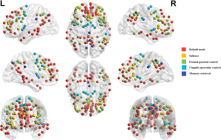
Methods: Network segregation, calculated from resting state functional magnetic resonance imaging scans, was measured in 32 patients with MDD who entered a sham-controlled, double-blinded, randomized trial of rTMS to the left dorsolateral prefrontal cortex, and a cohort of 20 healthy controls (HCs). Half of the MDD patients received sham treatment in the blinded phase, followed by active rTMS in the open-label phase. The analyses focused on segregation of the following networks: default mode (DMN), salience (SN), fronto-parietal (FPN), cingulo-opercular (CON), and memory retrieval (MRN).
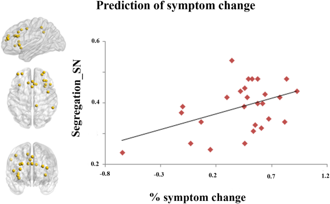
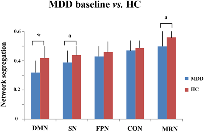
Results: There was no differential change in network segregation comparing sham to active treatment. However, in the combined group of patients who completed active rTMS treatment (in the blinded plus open-label phases), higher baseline segregation of SN significantly predicted more symptom improvement after rTMS. Compared to HCs at baseline, MDD patients showed decreased segregation in DMN, and trend-level decreases in SN and MRN.
Conclusion: The results highlight the importance of network segregation in MDD, particularly in the SN, where more normal baseline segregation of SN may predict better treatment response to rTMS in depression.
Keywords: Default mode network; Depression; Repetitive transcranial magnetic stimulation; Salience network; Segregation.
Copyright © 2019 The Authors. Published by Elsevier Inc. All rights reserved.
Figures
References
-
- Allen E.A., Erhardt E.B., Damaraju E., Gruner W., Segall J.M., Silva R.F., Havlicek M., Rachakonda S., Fries J., Kalyanam R., Michael A.M., Caprihan A., Turner J.A., Eichele T., Adelsheim S., Bryan A.D., Bustillo J., Clark V.P., Feldstein Ewing S.W., Filbey F., Ford C.C., Hutchison K., Jung R.E., Kiehl K.A., Kodituwakku P., Komesu Y.M., Mayer A.R., Pearlson G.D., Phillips J.P., Sadek J.R., Stevens M., Teuscher U., Thoma R.J., Calhoun V.D. A baseline for the multivariate comparison of resting-state networks. Front. Syst. Neurosci. 2011;5:2. - PMC - PubMed
-
- Bakker N., Shahab S., Giacobbe P., Blumberger D.M., Daskalakis Z.J., Kennedy S.H., Downar J. rTMS of the dorsomedial prefrontal cortex for major depression: safety, tolerability, effectiveness, and outcome predictors for 10 Hz versus intermittent theta-burst stimulation. Brain. Stimulation. 2015;8:208–215. - PubMed
-
- Carpenter L.L., Janicak P.G., Aaronson S.T., Boyadjis T., Brock D.G., Cook I.A., Dunner D.L., Lanocha K., Solvason H.B., Demitrack M.A. Transcranial magnetic stimulation (TMS) for major depression: a multisite, naturalistic, observational study of acute treatment outcomes in clinical practice. Depress. Anxiety. 2012;29:587–596. - PubMed
Publication types
MeSH terms
Grants and funding
LinkOut - more resources
Full Text Sources
