Review
. 2019 Feb 18;146(4):dev156059.
doi: 10.1242/dev.156059.

Neural stem cells: origin, heterogeneity and regulation in the adult mammalian brain

Affiliations
Review

Neural stem cells: origin, heterogeneity and regulation in the adult mammalian brain

Kirsten Obernier et al. Development. .

Abstract

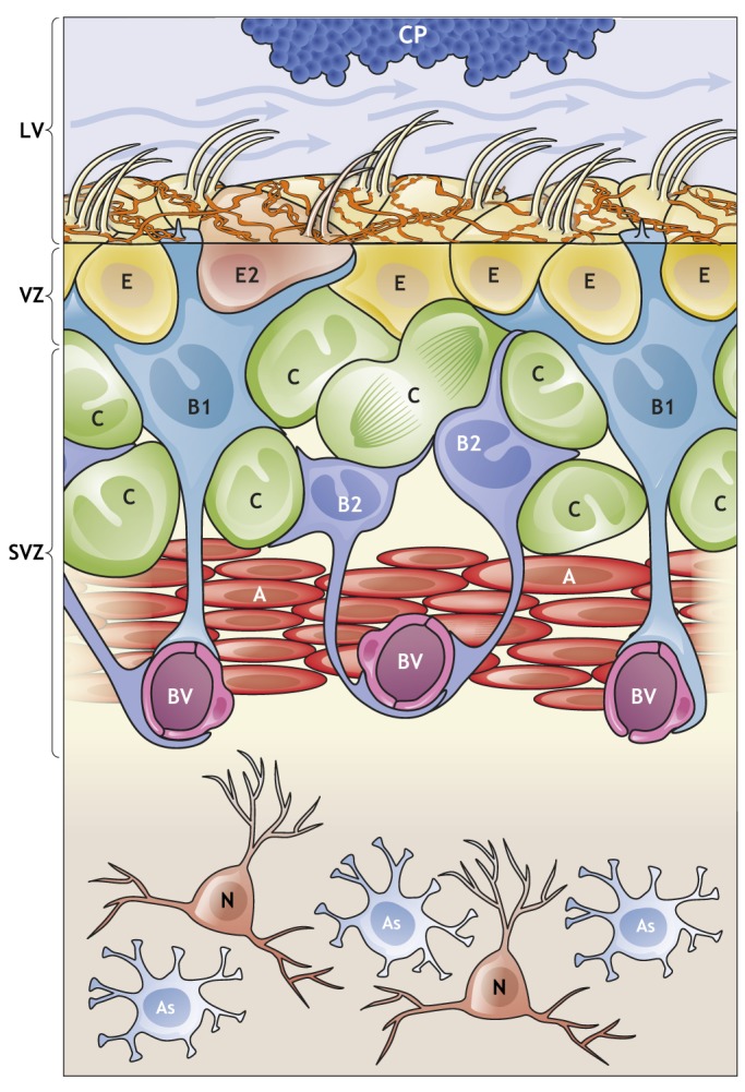
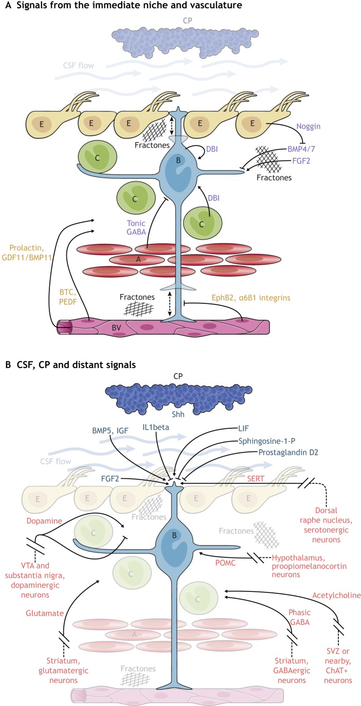
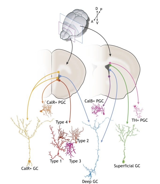
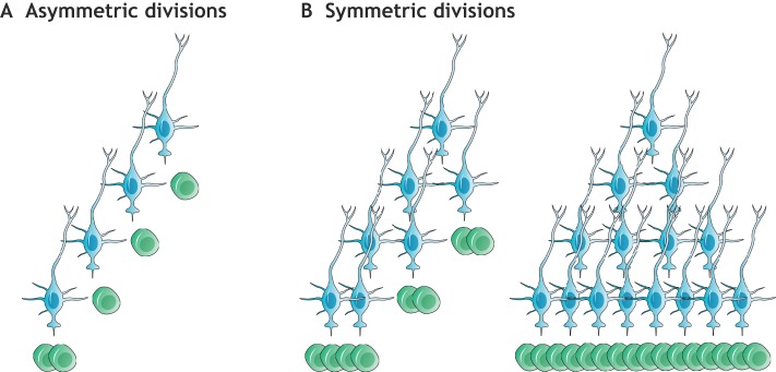
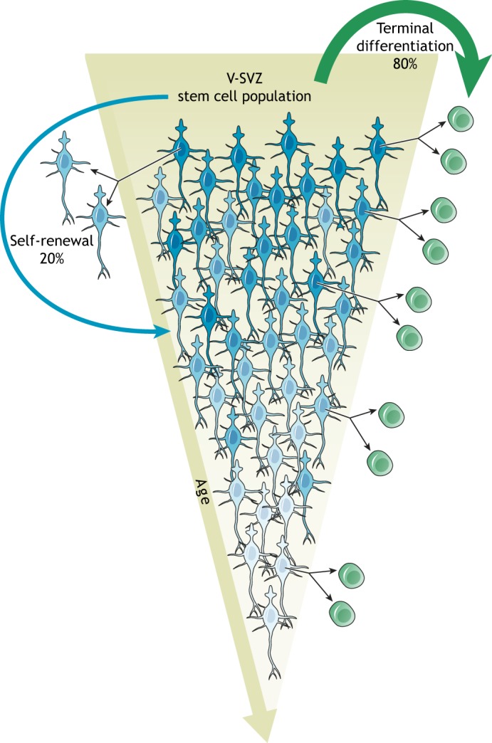
In the adult rodent brain, neural stem cells (NSCs) persist in the ventricular-subventricular zone (V-SVZ) and the subgranular zone (SGZ), which are specialized niches in which young neurons for the olfactory bulb (OB) and hippocampus, respectively, are generated. Recent studies have significantly modified earlier views on the mechanisms of NSC self-renewal and neurogenesis in the adult brain. Here, we discuss the molecular control, heterogeneity, regional specification and cell division modes of V-SVZ NSCs, and draw comparisons with NSCs in the SGZ. We highlight how V-SVZ NSCs are regulated by local signals from their immediate neighbors, as well as by neurotransmitters and factors that are secreted by distant neurons, the choroid plexus and vasculature. We also review recent advances in single cell RNA analyses that reveal the complexity of adult neurogenesis. These findings set the stage for a better understanding of adult neurogenesis, a process that one day may inspire new approaches to brain repair.

Keywords: Differentiation; Neural stem cells; Neurogenesis; Self-renewal; Stem cell heterogeneity; Transcriptomics.


Figures

Fig. 1.
Fig. 1.
The V-SVZ: a niche for adult neurogenesis. Schematic of the organization and composition of the adult V-SVZ in the walls of the lateral ventricle (LV). B1 cells (light blue) have astroglial characteristics and function as the NSCs in the V-SVZ. They give rise to B2 cells (dark blue), which share many astroglial characteristics with B1 cells, including contacts with blood vessels (BV), but lack an apical contact. B1 cells also generate transient-amplifying cells (C cells; green) that give rise to young neurons (A cells; red). B1 cells are in contact with the cerebrospinal fluid (CSF; blue arrows) through a small apical contact that harbors a primary cilium. The choroid plexus (CP; blue) secretes factors into the CSF that are important for the regulation of B1 cells (see Fig. 2); the CP and CSF are therefore included as part of the niche. B1 cell apical endings are surrounded by the large surfaces of multiciliated and biciliated ependymal cells (E/E2 cells) which form pinwheel-like structures. In addition, supraependymal axons (orange) coursing on the surface of the ventricular wall contact both E cells and B1 cells. Below the apical surface, in the SVZ, B1 cells contact C cells, A cells, BV and B2 cells. Mature neurons (N; orange) and astrocytes (As; pale blue) can be found below the V-SVZ in the striatum.
Fig. 2.
Fig. 2.
Signaling molecules that regulate V-SVZ neurogenesis. (A,B) A number of signaling molecules promote or inhibit the proliferation of B1 cells and hence regulate neurogenesis. These regulators can come from cells in direct contact with B1 cells (the ‘immediate’ niche) or from more distant sources. (A) Nearby cells, such as C cells, A cells, other B1 cells and E cells release factors (purple) that can influence B1 cells. B1 cells also contact blood vessels (BV) with the end foot of their basal process, which allows cell-cell interaction with endothelial cells and access to soluble factors (orange) in the blood. Dashed arrows indicate as yet unknown actions. (B) Factors (dark blue) from the CSF and CP act on B1 cells through their apical contacts. A number of distant signals (red), such as neurotransmitters released from neurons that reside outside the niche, also act on NSCs and their progeny.
Fig. 3.
Fig. 3.
NSC heterogeneity. B1 cells are regionally specified: depending on their location in the V-SVZ along the anterior-posterior and medial-lateral axes, B1 cells generate different subtypes of interneurons that are destined for the OB. For example, B1 cells in the posterior dorsal V-SVZ (indicated in the posterior coronal section on the right) generate tyrosine-hydroxylase (TH)+ periglomerular cells (PGCs; pink) and superficial granule cells (GCs; green), whereas B1 cells in the posterior ventral V-SVZ give rise to calbindin (CalB)+ PGCs (purple). Cells in the anterior medial V-SVZ (shown in the anterior section on the left) generate calretinin (CalR)+ PGCs (orange). Ventral NSCs produce deep GCs (blue), whereas dorsal NSCs generate superficial GCs. A small subset of NSCs in the lateral anterior ventral V-SVZ generates four additional types of interneurons (type 1-4).
Fig. 4.
Fig. 4.
Division modes of NSCs. (A) Asymmetric stem cell divisions (proposed e.g. by Calzolari et al., 2015) generate a fixed lineage with one B1 cell (blue) giving rise to another B1 cell and one C cell (green). This mode of division requires repeated divisions of B1 cells for the production of an increased population of progeny before a final (consuming) division generates only C cells. (B) Symmetric divisions can generate either two B1 cells (symmetric self-renewal) or two C cells (symmetric consumption/differentiation), allowing self-renewal and differentiation to be regulated independently, and allowing the generation of progeny with minimal replicative burden on individual stem cells. Note that sister B1 cells can become consumed asynchronously (left), and that multiple self-renewing divisions can precede consuming divisions (right).
Fig. 5.
Fig. 5.
B1 cells decline over time. B1 cells (blue) divide symmetrically. The majority (∼80%) of B1 cells becomes consumed by the generation of C cells (green), leading to a declining B1 cell population as the animal ages (terminal differentiation; green arrow). A smaller fraction (∼20%) of B1 cells symmetrically self-renews, which generates two B1 cells. Given the high level of terminal differentiation, this process allows neurogenesis to be maintained throughout the lifespan of the animal (albeit at lower levels). Note that B1 cells can undergo more than one round of symmetric self-renewal, enter extended periods of quiescence, and generate progeny at different times. In addition, each self-renewing division may lead to intrinsic changes in B1 cells (aging of the stem cell lineage), here illustrated by different shades of blue.

References

    1. Aguirre A., Rubio M. E. and Gallo V. (2010). Notch and EGFR pathway interaction regulates neural stem cell number and self-renewal. Nature 467, 323-327. 10.1038/nature09347 - DOI - PMC - PubMed
    1. Alfonso J., Le Magueresse C., Zuccotti A., Khodosevich K. and Monyer H. (2012). Diazepam binding inhibitor promotes progenitor proliferation in the postnatal SVZ by reducing GABA signaling. Cell Stem Cell 10, 76-87. 10.1016/j.stem.2011.11.011 - DOI - PubMed
    1. Altman J. (1962). Are new neurons formed in the brains of adult mammals? Science 135, 1127-1128. 10.1126/science.135.3509.1127 - DOI - PubMed
    1. Altman J. and Das G. D. (1965). Autoradiographic and histological evidence of postnatal hippocampal neurogenesis in rats. J. Comp. Neurol. 124, 319-335. 10.1002/cne.901240303 - DOI - PubMed
    1. Alvarez-Buylla A., Theelen M. and Nottebohm F. (1990). Proliferation “hot spots” in adult avian ventricular zone reveal radial cell division. Neuron 5, 101-109. 10.1016/0896-6273(90)90038-H - DOI - PubMed

Publication types

MeSH terms