. 2019 Apr 1;202(7):2153-2163.
doi: 10.4049/jimmunol.1801613. Epub 2019 Feb 18.

ARTC2.2/P2RX7 Signaling during Cell Isolation Distorts Function and Quantification of Tissue-Resident CD8+ T Cell and Invariant NKT Subsets

Affiliations

ARTC2.2/P2RX7 Signaling during Cell Isolation Distorts Function and Quantification of Tissue-Resident CD8+ T Cell and Invariant NKT Subsets

Henrique Borges da Silva et al. J Immunol. .

Abstract

Peripheral invariant NKT cells (iNKT) and CD8+ tissue-resident memory T cells (TRM) express high levels of the extracellular ATP receptor P2RX7 in mice. High extracellular ATP concentrations or NAD-mediated P2RX7 ribosylation by the enzyme ARTC2.2 can induce P2RX7 pore formation and cell death. Because both ATP and NAD are released during tissue preparation for analysis, cell death through these pathways may compromise the analysis of iNKT and CD8+ TRM Indeed, ARTC2.2 blockade enhanced recovery of viable liver iNKT and TRM The expression of ARTC2.2 and P2RX7 on distinct iNKT subsets and TRM is unclear, however, as is the impact of recovery from other nonlymphoid sites. In this study, we performed a comprehensive analysis of ARTC2.2 and P2RX7 expression in iNKT and CD8+ T cells in diverse tissues, at steady-state and after viral infection. NKT1 cells and CD8+ TRM express high levels of both ARTC2.2 and P2RX7 compared with NKT2, NKT17, and CD8+ circulating memory subsets. Using nanobody-mediated ARTC2.2 antagonism, we showed that ARTC2.2 blockade enhanced NKT1 and TRM recovery from nonlymphoid tissues during cell preparation. Moreover, blockade of this pathway was essential to preserve functionality, viability, and proliferation of both populations. We also showed that short-term direct P2RX7 blockade enhanced recovery of TRM, although to a lesser degree. In summary, our data show that short-term in vivo blockade of the ARTC2.2/P2RX7 axis permits much improved flow cytometry-based phenotyping and enumeration of murine iNKT and TRM from nonlymphoid tissues, and it represents a crucial step for functional studies of these populations.


Figures

Fig.1.
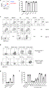
Fig.1.. Expression of ARTC2.2 and P2RX7 in iNKT and CD8+ T cell subsets.
(a) Representative flow cytometry plot showing expression of ARTC2.2 and P2RX7 or isotype control in liver iNKT cells. (b) Representative plots showing expression of ARTC2.2 and P2RX7 in NKT1, NKT2, or NKT17 cell subsets from the indicated tissues. N/A means not analyzed (due to paucity of cells of that subset in the tissue) (c) Percentage of NKT1 cells that are ARTC2.2+P2RX7+ (as defined by the quadrants in (a)). (d) Expression of ARTC2.2 and P2RX7 in polyclonal CD8+ T cells from the indicated tissues (above) and in virus specific CD8+ T cells 4 weeks after infection (below). For the virus specific CD8 T cell analysis, P14 CD8 T cells were adoptively transferred into congenic recipient mice (2.5 × 10⁴ cells/mouse), followed by infection with LCMV-Armstrong (2 × 10⁵ PFU, i.p.). After 4 weeks, cells were isolated from the indicated tissues. (e) The percentages of ARTC2.2+P2RX7+ cells are shown for polyclonal CD8+ T cells in the thymus and spleen, as well as splenic memory P14 cell subsets (central memory – TCM, effector memory – TEM, spleen resident memory – SLO TRM). (f) Percentages of memory P14 cells that were ARTC2.2+P2RX7+ in the different organs is shown, as well as polyclonal small intestine-resident CD8+ T cells. (a-f) Data are from 3 independent experiments, n=5–6 per experimental group.
Fig. 2.
Fig. 2.. Blockade of ARTC2.2/P2RX7 axis improves recovery of NKT1 cells in non-lymphoid tissues
Number of NKT1 cells recovered in the indicated tissues from mice treated with anti-ARTC2.2 nanobody or PBS prior to tissue harvest. NKT1 cells are defined as TCRβ+ CD1d tetramer+ T-bet+. Blue bar represents cells from mice received PBS, red bar represents cells from mice received anti-ARTC2.2 nanobody. mLN=mesenteric lymph node; LPL=lamina propria; IEL=intestinal epithelial lymphocyte. Data are from 8 independent experiments with 2–6 mice in each experiment.
Fig.3.
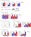
Fig.3.. Nanobody-mediated ARTC2.2 blockade enhances recovery of diverse TRM cells in tissue processing.
(a-e) P14 CD8 T cells were isolated from the indicated tissues, with injection of ARTC2.2 blocking nanobodies (Nanobody) or PBS. (a) P14 cell numbers per organ comparing the PBS and Nanobody-treated groups. (b) Side-by-side comparison between the fold differences observed in the results from (a), and the fold differences between flow cytometry vs quantitative immunofluorescence (QIM) observed by Steinert et al. (2015). (c) Representative flow cytometry plots showing expression of CD69 and CD103 in SI LPL P14 cells from PBS and Nanobody-treated mice. (d) Numbers of CD103+ and CD103 P14 cells from small intestines from PBS and Nanobody-treated mice. (e) Representative flow cytometry plots showing the percentages of CD8α i.v.+ (non-resident) vs i.v. (resident) P14 cells in the lungs of PBS and Nanobody-treated mice. (f-g) Mice were transferred with equal numbers of WT and P2rx7−/− P14 cells (identified by distinct congenic markers), followed by LCMV-Arm infection. (f) Representative flow cytometry plots of SI IEL P14 cells from Nanobody- or PBS-treated, co-transferred mice. (g) Numbers of WT (filled bars) and P2rx7−/− (open bars) P14 cells in the SI IEL of co-transferred mice, with PBS or Nanobody treatment. Data are from 3–4 independent experiments, n=6–15 per experimental group.
Fig. 4.
Fig. 4.. Blockade of ARTC2.2/P2RX7 axis preserves surface molecules, cytokine production and viability of iNKT cells
(a) Representative flow cytometry plots for surface expression of CD27, CD69, P2RX7, ARTC2.2, CD122 and CD4 in spleen and liver NKT1 cells from mice treated with anti-ARTC2.2 nanobody or PBS. (b-d) Liver mononuclear cells isolated from mice treated with anti-ARTC2.2 nanobody or PBS, were stimulated in vitro with presence of PMA/Ionomycin for 4 hours. (b) Numbers (left column) and frequency of NKT1 cells among total T cells (TCRβ+) (right column) after in vitro stimulation. (c) Representative flow cytometry plots for IFN-γ production in NKT1 cells after in vitro stimulation. (d) Frequency (left column) and number (right column) of IFN-γ+ NKT1 cells after in vitro stimulation. (e-f) Liver mononuclear cells isolated from mice treated with anti-ARTC2.2 nanobody or PBS, were cultured in vitro for 24 or 72 hours. (e) Frequency of live iNKT cells (viability dye negative) after in vitro culture for 24 hours (upper row) or 72 hours (bottom row). (f) Frequency (left column) and number (right column) of live iNKT cells after in vitro culture for 24 hours (upper row) or 72 hours (bottom row). Data are from 4 independent experiments, n=2–10 mice per experiment.
Fig. 5.
Fig. 5.. Pre-harvest ARTC2.2 blockade preserves IEL TRM functions and viability during in vitro procedures.
(a) Median fluorescence intensity (MFI) values for ARTC2.2, P2RX7, CD69 and CD44 in liver P14 cells from mice treated with Nanobody or PBS. (b) Experimental design for the in vitro experiments done with spleen and SI IEL P14 cells. (c) Percentages (left) and numbers/well (right) of PBS vs Nanobody-treated SI IEL P14 cells after 4h of in vitro culture without further stimulation (RPMI), PMA/Ionomycin, or gp33 peptide. (d) Representative flow cytometry plots showing IFN-γ production in SI IEL P14 cells from PBS and Nanobody-treated mice, stimulated in vitro with PMA/Ionomycin (4h). (e) Percentages (left) and numbers/well (right) of IFN-γ+ SI IEL P14 cells after 4h of in vitro culture without stimulation (RPMI), PMA/Ionomycin or gp33 peptide. (f) Percentages (left) and numbers/well (center), as well as the numbers of IFN-γ+/well (right) P14 cells after 24h of in vitro culture without any stimulation (RPMI) or with αCD3/αCD28. Data are from 2–4 independent experiments, n=4–13 per experimental group.
Fig. 6.
Fig. 6.. Short-term P2RX7 pharmacological blockade is an alternative for rescuing TRM cells during ex vivo procedures.
(a-b) P14 memory cells were isolated from the small intestine of mice after treatment with PBS, Brilliant Blue G (BBG) or A-438079. (a) Numbers of i.v. (resident) small intestine P14 cells in mice treated with PBS, BBG or A-438079. (b) Representative flow cytometry plots showing CD69 and CD103 expression in SI LPL P14 cells from mice treated with PBS, BBG or A-438079. (c-d) Mice were injected prior to sacrifice with PBS, A-438079 or Nanobody, and organs harvested and processed as described before. (c) Mitotracker green (MTG) median fluorescence averages of spleen (left) and SI IEL (right) P14 cells from mice treated with PBS or A-438079. (d) MTG (left) and Tetramethylrhodamine (TMRE) (right, normalized to MTG) median fluorescence averages of SI IEL P14 cells from mice treated with PBS or ARTC2.2 Nanobody. Data are from 2–3 independent experiments, n=3–10 per experimental group.

Similar articles

Cited by

References

    1. Scheuplein F, Schwarz N, Adriouch S, Krebs C, Bannas P, Rissiek B, Seman M, Haag F, and Koch-Nolte F. 2009. NAD+ and ATP released from injured cells induce P2X7-dependent shedding of CD62L and externalization of phosphatidylserine by murine T cells. J Immunol 182: 2898–2908. - PubMed
    1. Di Virgilio F, Dal Ben D, Sarti AC, Giuliani AL, and Falzoni S. 2017. The P2X7 Receptor in Infection and Inflammation. Immunity 47: 15–31. - PubMed
    1. Bartlett R, Stokes L, and Sluyter R. 2014. The P2X7 receptor channel: recent developments and the use of P2X7 antagonists in models of disease. Pharmacol Rev 66: 638–675. - PubMed
    1. Kawamura H, Aswad F, Minagawa M, Govindarajan S, and Dennert G. 2006. P2X7 receptors regulate NKT cells in autoimmune hepatitis. J Immunol 176: 2152–2160. - PubMed
    1. Proietti M, Cornacchione V, Rezzonico Jost T, Romagnani A, Faliti CE, Perruzza L, Rigoni R, Radaelli E, Caprioli F, Preziuso S, Brannetti B, Thelen M, McCoy KD, Slack E, Traggiai E, and Grassi F. 2014. ATP-gated ionotropic P2X7 receptor controls follicular T helper cell numbers in Peyer’s patches to promote host-microbiota mutualism. Immunity 41: 789–801. - PubMed

Publication types

MeSH terms