

Profile of childhood glaucoma at a tertiary center in South India
- PMID: 30777953
- PMCID: PMC6407385
- DOI: 10.4103/ijo.IJO_786_18
Profile of childhood glaucoma at a tertiary center in South India
Abstract
Purpose: To describe the prevalence of various types of childhood glaucomas, their clinical features and treatment methods.
Methods: We prospectively included consecutive children with glaucoma presenting to glaucoma clinic for the first time between March 2013 and May 2014. We classified childhood glaucomas as per the classification proposed by Congenital Glaucoma Research Network.
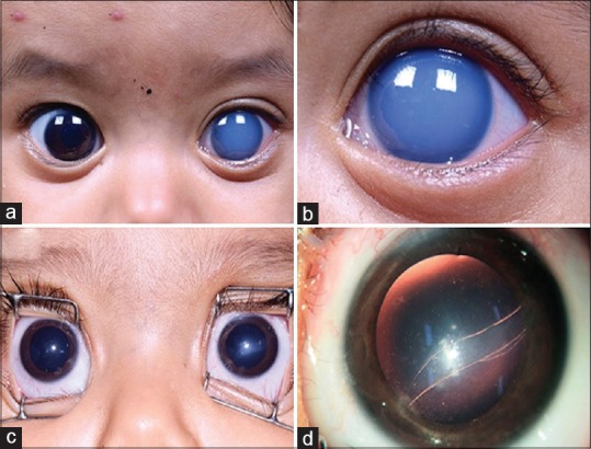
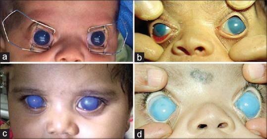
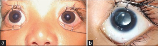
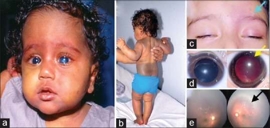
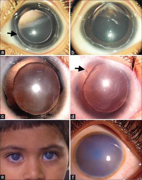
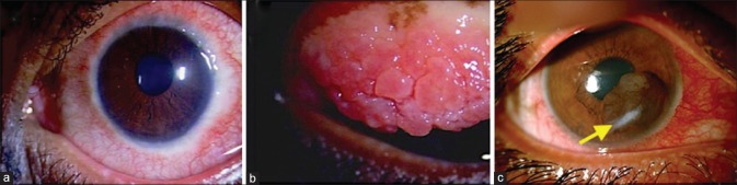
Results: Of the 275 children (449 eyes) with glaucoma during this period, primary glaucomas constituted 56% (n = 252 eyes of 145 children), including 169 eyes (37.64%) of 97 children with primary congenital glaucoma (PCG), 16 eyes (3.56%) of 10 children with infantile glaucoma, and 67 eyes (14.9%) of 38 children with juvenile open angle glaucoma. Among these, 85% (214 eyes of 107 children) had bilateral involvement. Secondary glaucomas constituted 44%; they were glaucoma associated with ocular anomalies 18% (n = 80 eyes), glaucoma associated with acquired conditions (steroid induced and traumatic glaucoma) 14% (n = 61 eyes), glaucoma following congenital cataract surgery 7.6% (n = 34 eyes), and glaucoma associated with systemic or syndromic conditions 5% (n = 22 eyes). In primary glaucomas, boys and girls were equally affected (1:1), and more boys (3.8:1) had acquired glaucomas. Close to 80% PCG eyes were managed surgically with combined trabeculotomy and trabeculectomy (CTT). Majority of secondary glaucomas were managed medically.
Conclusion: In our cohort, PCG was the most common childhood glaucoma and CTT was the most common surgery performed in these eyes. Steroid-induced and traumatic glaucomas were the most common acquired glaucomas; appropriate measures should be taken to avert these preventable glaucomas in children.
Keywords: Childhood glaucoma in India; South India; childhood glaucomas; congenital glaucoma; prevalence of glaucoma in children; profile of glaucoma in children.
Conflict of interest statement
None
Figures
References
-
- Gilbert CE, Rahi JS, Quinn GE. 2 ed. London: Edward Arnold Ltd; 2003. Visual Impairment and Blindness in Children.
-
- Gencik A. Epidemiology and genetics of primary congenital glaucoma in Slovakia. Description of a form of primary congenital glaucoma in gypsies with autosomal-recessive inheritance and complete penetrance. Dev Ophthalmol. 1989;16:76–115. - PubMed
-
- Sarfarazi M, Stoilov I. Molecular genetics of primary congenital glaucoma. Eye (Lond) 2000;14:422–8. - PubMed
MeSH terms
LinkOut - more resources
Full Text Sources
Medical
