. 2019 Feb 18;10(1):798.
doi: 10.1038/s41467-019-08739-4.

HIV-1 vaccination by needle-free oral injection induces strong mucosal immunity and protects against SHIV challenge

Affiliations

HIV-1 vaccination by needle-free oral injection induces strong mucosal immunity and protects against SHIV challenge

Andrew T Jones et al. Nat Commun. .

Abstract

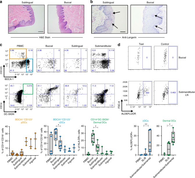
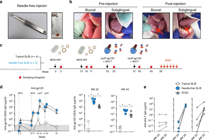
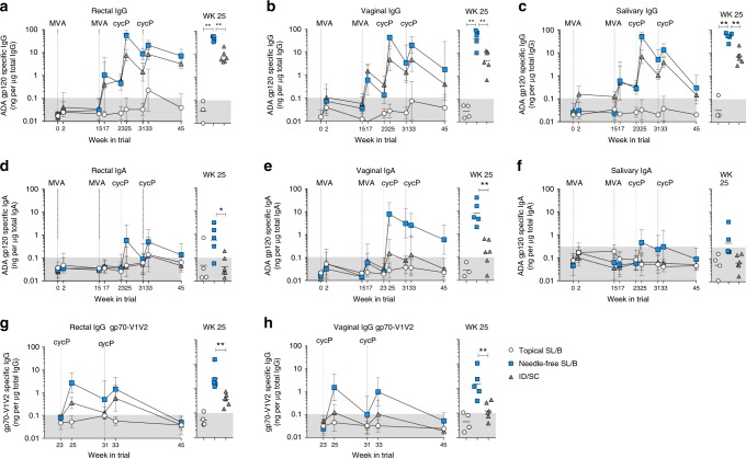
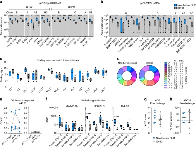
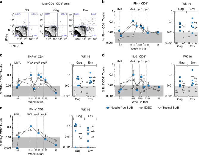
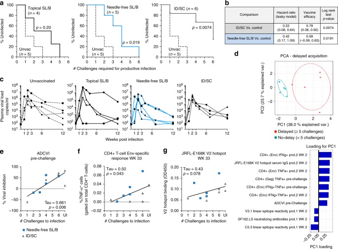
The oral mucosa is an attractive site for mucosal vaccination, however the thick squamous epithelium limits antigen uptake. Here we utilize a modified needle-free injector to deliver immunizations to the sublingual and buccal (SL/B) tissue of rhesus macaques. Needle-free SL/B vaccination with modified vaccinia Ankara (MVA) and a recombinant trimeric gp120 protein generates strong vaccine-specific IgG responses in serum as well as vaginal, rectal and salivary secretions. Vaccine-induced IgG responses show a remarkable breadth against gp70-V1V2 sequences from multiple clades of HIV-1. In contrast, topical SL/B immunizations generates minimal IgG responses. Following six intrarectal pathogenic SHIV-SF162P3 challenges, needle-free but not topical immunization results in a significant delay of acquisition of infection. Delay of infection correlates with non-neutralizing antibody effector function, Env-specific CD4+ T-cell responses, and gp120 V2 loop specific antibodies. These results demonstrate needle-free MVA/gp120 oral vaccination as a practical and effective route to induce protective immunity against HIV-1.


Conflict of interest statement

The authors declare no competing interests.

Figures

Fig. 1
Fig. 1
Dendritic cells in the sublingual and buccal (SL/B) Tissue of rhesus macaques. Paraffin-embedded sublingual and buccal tissue sections were stained with a Hematoxylin and eosin and imaged by light microscopy (scale bar, 500 μm) or b anti-Langerin and analyzed by immunohistochemistry (scale bar, 200 μm). Arrows show Langerin+ cells stained in red. c Representative flow plots showing dendritic cell (DC) subsets in PBMC, sublingual tissue, buccal tissue, and the submandibular, submental, and inguinal lymph nodes. Top row; CD45+CD3CD20HLA-DR+CD14CD16 cells were gated for plasmacytoid DC (CD123+BDCA-1, orange) and conventional DC (CD123BDCA-1+, blue) markers. Middle row; CD45+CD3CD20HLA-DR+ cells were gated for Dermal DCs (CD14+DC-SIGN+, green). Bottom row; frequencies of DC subsets in different tissues (*, p < 0.05, Wilcoxon matched-pairs test). Data representative of samplings from six independent animals, n = 5 (PBMC) n = 6 (buccal, sublingual), n = 3 (submandibular, submental, and inguinal lymph nodes). Samples from the same animal were paired together. d Detection of aldehyde dehydrogenase (ALDH) activity in buccal or submandibular DCs using the ALDEFLUORTM fluorescent reagent. Control samples were used to set gating. Representative flow plots are shown on top measuring ALDH activity in conventional DCs in buccal tissue compared to submandibular lymph nodes. Bottom, frequencies of ALDEFLUOR+ conventional DCs (blue) and dermal DCs (green) in PBMCs (n = 2), buccal (n = 3) and sublingual tissue (n = 3), and submandibular (n = 3) and submental lymph nodes (n = 3) (*, p < 0.05; **, p < 0.01; Mann–Whitney test). PMBC, peripheral blood mononuclear cells; SL/B, sublingual and buccal tissue. Box and whiskers plot in c, d; box extends from 25th to 75th percentile, the line indicates median, whiskers indicate min and max values
Fig. 2
Fig. 2
SL/B immunization with a needle-free injector induces strong systemic antibody responses. a Syrijet, the needle-free injector used to deliver immunizations to the sublingual and buccal tissue. Sterile water cartridges were modified to contain immunogens. b Sublingual and buccal tissue of a rhesus macaque before and five minutes after 100 μl PBS injection via Syrijet. c Study design. Rhesus macaques (n = 15) were immunized twice with MVA-HIV (1 × 108 pfu) and boosted twice with recombinant trimeric gp120 (cycP-gp120) (100 μg) with the mucosal adjuvant dmLT. Animals were immunized via topical application to the sublingual and buccal (SL/B) tissue (n = 4), needle-free injection to the SL/B tissue (n = 5), or intradermally (with MVA-HIV) and subcutaneously (with cycP-gp120 + dmLT) (n = 6). MVA-HIV and cycP-gp120 doses were split between the buccal and sublingual tissue (SL/B) or the left and right thigh (ID/SC). 19 weeks following the second cycP-gp120 immunization, animals were challenged intra-rectally with low dose pathogenic SHIV-SF162P3 weekly for up to six weeks. Cartoons depict MVA-HIV and virus-like particles, cycP-gp120, and dmLT. d Kinetics of anti-gp120 (ADA) serum IgG in vaccine groups (geomean ± SD) and for individual animals (line, geomean ± SD) at the peak time point (wk 25) and pre-challenge time point (wk 45) (*, p < 0.05; Mann–Whitney test). Dotted lines denote week of indicated immunization. e Anti-dmLT serum IgG response in animals before (wk 25) and two weeks post (wk 45) immunization with cycP-gp120 + dmLT. d, e White circle, topical SL/B (n = 4); blue square, needle-free SL/B (n = 5); gray triangle, ID/SC (n = 6)
Fig. 3
Fig. 3
Needle-free SL/B immunization generates strong mucosal antibody responses. ac Anti-ADA gp120 IgG antibodies in rectal (a), vaginal (b), and salivary (c) secretions. df Anti-ADA gp120 IgA antibodies in rectal (d), vaginal (e), and salivary (f) secretions. Data represented as geomean ± SD specific activity for each group. Specific activity calculated as ng gp120-specific IgG or IgA antibody per μg total IgG or IgA isolated. Specific activity for individual animals is shown at the peak time point (wk 25); line indicates geomean. The shaded region in each graph indicates the specific activity cut-off value. gh IgG specific activity against gp70-V1V2 (Clade B/Case A2) in rectal and vaginal secretions. ah * p < 0.05; ** p < 0.01; Mann–Whitney test. White circle, topical SL/B (n = 4); blue square, needle-free SL/B (n = 5); gray triangle, ID/SC (n = 6)
Fig. 4
Fig. 4
Serum IgG specificity, neutralizing activity, and effector function. Binding of IgG antibodies at two weeks post the first cycP-gp120 immunization (wk 25) to a gp120 and gp140 antigens and b gp70-V1V2 scaffolds representing the global diversity of HIV-1 determined using Binding Antibody Multiplex Assay (BAMA). Shaded areas indicate clade. c Binding of peak immune sera (wk 25) to 15mer peptides derived from clade B consensus gp120, measured by peptide microarray linear epitope mapping and reported as binding signal (Log2 fold difference post-immunization/baseline binding intensity). Magnitude of binding to each epitope is defined as the highest binding signal for a single peptide within the region of the epitope. d Representation of peptide array binding of each clade B consensus epitope as % of the total response. e Anti-V2 hotspot (HS) response (wk 25) against V2-HS peptides derived from strains JRFL (E168K), ADA, and SF162P3 measured by ELISA. Dotted lines connect data from same animal. Blue squares, needle-free SL/B; gray triangles, ID/SC. f Neutralizing antibodies over time against HIV-1 isolates MW965.26, SF162.LS, and BaL.26, measured as ID50. g Antibody-dependent phagocytosis (ADP) at pre-challenge (wk 45). ADP score (mean ± S.D.) calculated for each serum by dividing the median fluorescence intensity (MFI) of bead positive cells by the value obtained using the same dilution of pooled serum from naive macaques. h Antibody-dependent cell viral inhibition (ADCVI) measured at pre-challenge as % viral inhibition (mean ± S.D.). An average of two individual experiments is shown. Box and whiskers plots (ac, f); box extends from 25th to 75th percentile, line indicates median, whiskers indicate min and max values (*p < 0.05; **p < 0.01; ***p < 0.001; ****, p < 0.0001; 2-Way ANOVA, multiple comparisons). ID50, serum dilution required to neutralize 50% infection
Fig. 5
Fig. 5
Needle-free SL/B immunization generates vaccine-specific CD4 and CD8 T cells in the blood. PMBCs were stimulated with HIV-1 consensus B Gag and Env peptides and analyzed by flow cytometry for cytokine production. a Representative flow plots for IFN-γ and TNF-α cytokine expression on Live CD3+CD4+ cells in non-stimulated (NS), Gag, or Env stimulated PBMCs is shown. Kinetics of the total (Gag + Env) b IFN-γ, c TNF-α, and d IL-2 response in CD4+ T cells (mean ± S.D.), with the peak response (wk 16) highlighted for each animal (line denotes mean). e Kinetics of the total IFN-γ response in CD8+ cells (mean ± S.D.), with the peak response (wk 16) highlighted for each animal. White circle, topical SL/B (n = 4); blue square, needle-free SL/B (n = 5); gray triangle, ID/SC (n = 6)
Fig. 6
Fig. 6
Needle-Free SL/B and ID/SC immunization results in delayed acquisition of SHIV-SF162P3 infection. Animals were challenged weekly with an intra-rectal low dose (1:100 dilution) of SHIV-SF162P3. 5 unvaccinated macaques were used as a control. a Acquisition of infection in topical SL/B, needle-free SL/B, or ID/SC immunized animals (Log-rank (Mantel-Cox) test). Dotted line, unvaccinated controls (n = 5); black line, topical SL/B (n = 4); blue line, needle-free SL/B (n = 5); gray line, ID/SC (n = 6). b Vaccine efficacy of ID/SC, needle-free SL/B immunization groups vs control animals (Log-rank (Mantel-Cox) test). c Kinetics of plasma viral loads in unvaccinated and vaccinated animals. d Top, Principal Comparison Analysis (PCA) plot showing PC1 and PC2 scores for delayed (≥5 challenges to infect, n = 5) or non-delayed (<5 challenges to infect, n = 6) acquisition of infection in needle-free SL/B and ID/SC immunized animals. Bottom, loadings of immune response parameters in PC1. e-g Correlation analysis of ADCVI activity at pre-challenge, Env-specific CD4+ T cell TNF-α production two weeks after the second cycP-gp120 immunization (wk 33), and JRFL-E168K V2 hotspot reactivity in serum IgG (wk 33) with acquisition of infection. (Kendall’s Tau Correlation Test). Needle-free SL/B (blue squares) and ID/SC (gray triangles) are combined for analysis (n = 11)

Similar articles

Cited by

References

    1. Shaw, G. M. & Hunter, E. HIV transmission. Cold Spring Harb. Perspect. Med. 2, 10.1101/cshperspect.a006965 (2012). - PMC - PubMed
    1. Haase AT. Early events in sexual transmission of HIV and SIV and opportunities for interventions. Annu. Rev. Med. 2011;62:127–139. doi: 10.1146/annurev-med-080709-124959. - DOI - PubMed
    1. Moir S, Chun TW, Fauci AS. Pathogenic mechanisms of HIV disease. Annu. Rev. Pathol. 2011;6:223–248. doi: 10.1146/annurev-pathol-011110-130254. - DOI - PubMed
    1. Lycke N. Recent progress in mucosal vaccine development: potential and limitations. Nat. Rev. Immunol. 2012;12:592–605. doi: 10.1038/nri3251. - DOI - PubMed
    1. Tuero I, Robert-Guroff M. Challenges in mucosal HIV vaccine development: lessons from non-human primate models. Viruses. 2014;6:3129–3158. doi: 10.3390/v6083129. - DOI - PMC - PubMed

Publication types

MeSH terms