. 2019 Feb 18;9(1):2205.
doi: 10.1038/s41598-018-38257-0.

Three-Dimensional Mass Spectrometry Imaging Identifies Lipid Markers of Medulloblastoma Metastasis

Affiliations

Three-Dimensional Mass Spectrometry Imaging Identifies Lipid Markers of Medulloblastoma Metastasis

Martin R L Paine et al. Sci Rep. .

Abstract

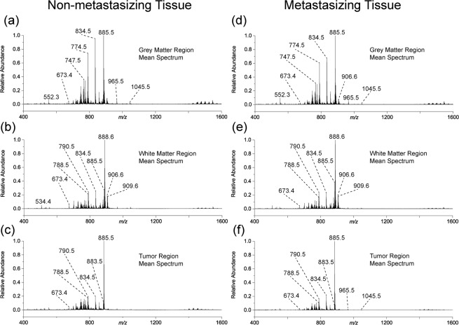
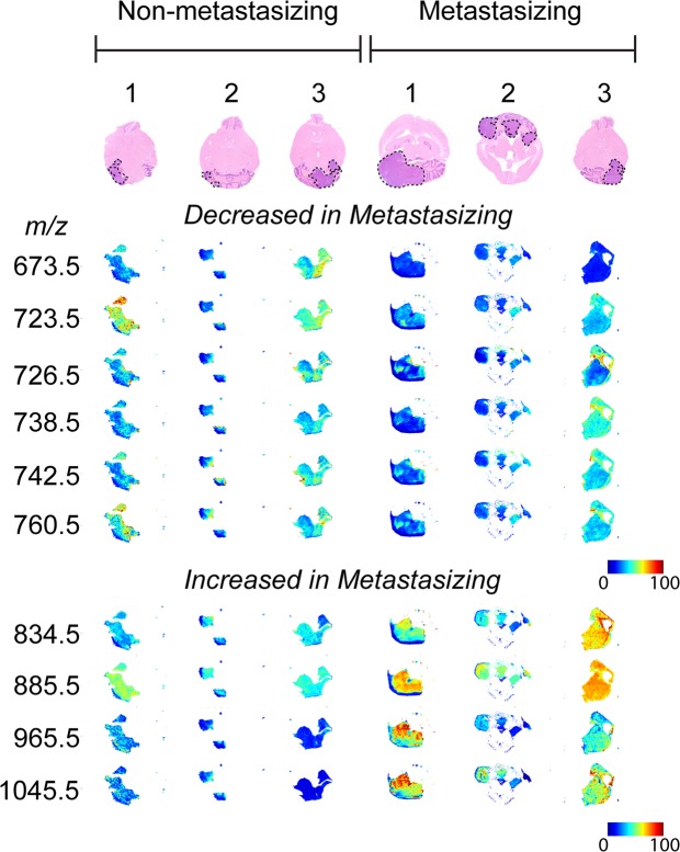
Treatment for medulloblastoma (MB) - the most common malignant pediatric brain tumor - includes prophylactic radiation administered to the entire brain and spine due to the high incidence of metastasis to the central nervous system. However, the majority of long-term survivors are left with permanent and debilitating neurocognitive impairments as a result of this therapy, while the remaining 30-40% of patients relapse with terminal metastatic disease. Development of more effective targeted therapies has been hindered by our lack of understanding of the underlying mechanisms regulating the metastatic process in this disease. To understand the mechanism by which MB metastasis occurs, three-dimensional matrix-assisted laser desorption/ionization mass spectrometry imaging (MALDI-MSI) experiments were performed on whole brains from a mouse model of human medulloblastoma. Analyzing the tumor and surrounding normal brain in its entirety enabled the detection of low abundance, spatially-heterogeneous lipids associated with tumor development. Boundaries of metastasizing and non-metastasizing primary tumors were readily defined, leading to the identification of lipids associated with medulloblastoma metastasis, including phosphatidic acids, phosphatidylethanolamines, phosphatidylserines, and phosphoinositides. These lipids provide a greater insight into the metastatic process and may ultimately lead to the discovery of biomarkers and novel targets for the diagnosis and treatment of metastasizing MB in humans.


Conflict of interest statement

J.H.K. and D.T. are employed by SCiLS Lab, proprietors of the software platform used in this study. M.R.L.P., S.R.E., J.L., D.H., T.J.M., R.M.A.H., and F.M.F. declare no potential conflict of interest.

Figures

Figure 1
Figure 1
(a) Optical images of the 49 aligned sagittal sections from a ND2:SmoA1 transgenic mouse brain containing a non-metastasizing cerebellum tumor with (b) the corresponding MS visualization using three representative channels; m/z 790.5 in blue, m/z 888.6 in green, and m/z 885.5 in red, overlaid on one another. (c) Hematoxylin and eosin (H&E) stains of sections no. 29 and 41 indicating the primary MB tumor region (purple). (d) Three-dimensional alignment of all 49 mouse brain sections shown in (b). (e–k) Visualization of two sagittal (e,f), three coronal (g–i), and two transversal (j,k) virtual sections.
Figure 2
Figure 2
(a) The bisecting k-means segmentation of 49 sagittal tissue sections from a ND2:SmoA1 transgenic mouse brain containing a non-metastasizing cerebellum tumor showing clear delineation of grey matter (blue), white matter (red), and the primary tumor (yellow). (b) Visualization of the k-means segmentation map in a reconstructed 3D volume as a transverse virtual section.
Figure 3
Figure 3
The calculated mean spectra for (a) grey matter, (b) white matter, and (c) tumor regions mouse brain containing a non-metastasizing medulloblastoma primary tumor and the (d) grey matter, (e) white matter, and (f) tumor regions in a mouse brain containing a metastasizing medulloblastoma primary tumor as defined by k-means segmentation. The lipidome of the tumor is comparatively very different to the grey and white matter regions based on changes in the measured relative abundances of metabolites throughout the spectrum. Differences between metastasizing and non-metastasizing tumors are subtler.
Figure 4
Figure 4
Optical images of a single transverse tissue section following H&E staining (tumor regions defined by black dotted line) for each of the six mouse brains used in the comparative study and the corresponding extracted-ion images for the ten lipids identified in Table 1 shown as 2-dimensional false-color plots. For each m/z values visualized distribution, TIC normalization was applied across all six tumor regions for direct comparison. The extracted-ion images highlight the heterogeneous spatial distribution within each tumor region and also general comparative changes across the six individual tumors.

References

    1. Bennett, J. et al. The clinical significance of equivocal findings on spinal MRI in children with medulloblastoma. Pediatr. Blood Cancer64 (2017). - PubMed
    1. Brinkman TM, et al. Cerebral white matter integrity and executive function in adult survivors of childhood medulloblastoma. Neuro Oncol. 2012;14:25–36. doi: 10.1093/neuonc/nos214. - DOI - PMC - PubMed
    1. Schwalbe EC, et al. Novel molecular subgroups for clinical classification and outcome prediction in childhood medulloblastoma: a cohort study. Lancet Oncol. 2017;18:958–971. doi: 10.1016/S1470-2045(17)30243-7. - DOI - PMC - PubMed
    1. Ramaswamy V, et al. Risk stratification of childhood medulloblastoma in the molecular era: the current consensus. Acta Neuropathol. 2016;131:821–831. doi: 10.1007/s00401-016-1569-6. - DOI - PMC - PubMed
    1. Leber MF, Efferth T. Molecular principles of cancer invasion and metastasis (review) Int. J. Oncol. 2009;34:881–895. - PubMed

Publication types