. 2019 Feb 18;9(1):2198.
doi: 10.1038/s41598-019-38651-2.

Delayed Captopril Administration Mitigates Hematopoietic Injury in a Murine Model of Total Body Irradiation

Affiliations

Delayed Captopril Administration Mitigates Hematopoietic Injury in a Murine Model of Total Body Irradiation

Elizabeth A McCart et al. Sci Rep. .

Abstract

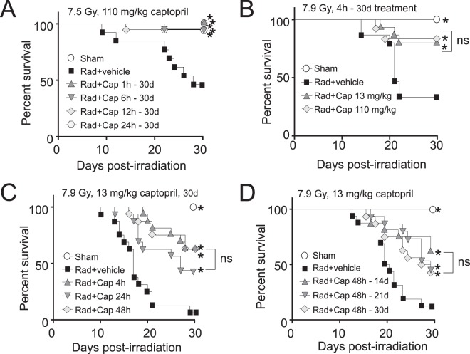
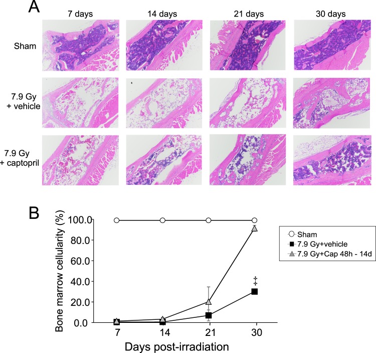
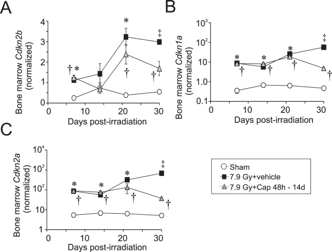
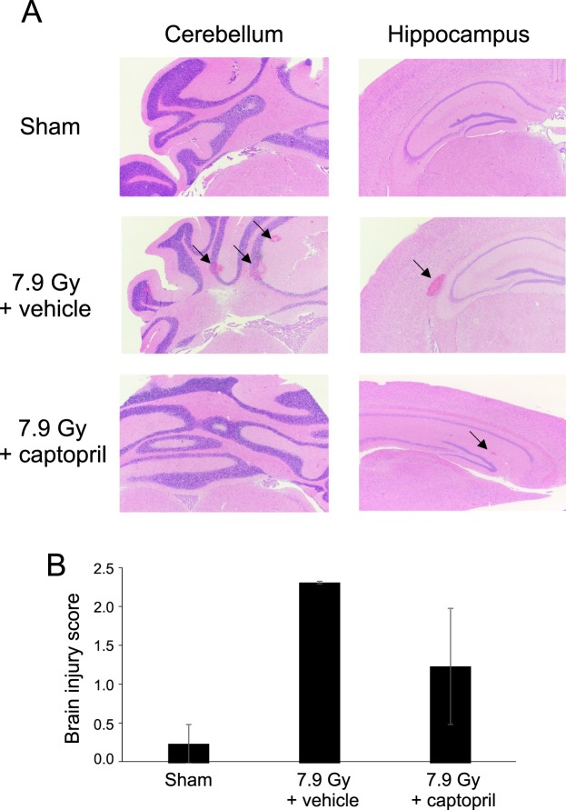
The increasing potential for accidental radiation exposure from either nuclear accidents or terrorist activities has escalated the need for radiation countermeasure development. We previously showed that a 30-day course of high-dose captopril, an ACE inhibitor, initiated 1-4 h after total body irradiation (TBI), improved Hematopoietic Acute Radiation Syndrome (H-ARS) and increased survival in mice. However, because of the time likely required for the deployment of a stockpiled radiation countermeasure to a radiation mass casualty site, there is a need for therapies that can be administered 24-48 hours after initial exposure. Using C57BL/6 mice exposed to an LD50-80/30 of 60Co TBI (7.75-7.9 Gy, 0.615 Gy/min), we show that low-dose captopril administration, initiated as late as 48 h post-TBI and continued for 14 days, significantly enhanced overall survival similarly to high-dose, rapid administration. Captopril treatment did not affect radiation-induced cell cycle arrest genes or the immediate loss of hematopoietic precursors. Reduced mortality was associated with the recovery of bone marrow cellularity and mature blood cell recovery at 21-30 days post-irradiation. Captopril reduced radiation-induced cytokines EPO, G-CSF, and SAA in the plasma. Finally, delayed captopril administration mitigated brain micro-hemorrhage at 21 days post-irradiation. These data indicate that low dose captopril administered as late as 48 h post-TBI for only two weeks improves survival that is associated with hematopoietic recovery and reduced inflammatory response. These data suggest that captopril may be an ideal countermeasure to mitigate H-ARS following accidental radiation exposure.


Conflict of interest statement

The authors declare no competing interests.

Figures

Figure 1
Figure 1
Kaplan-Meier Curves of the effects of reduced dosage and delayed administration of captopril on survival from total body irradiation. C57BL/6 mice, 12–14 weeks of age, were exposed to total body 60Co irradiation (0.6 Gy/min) or sham irradiated (sham). Irradiated (Rad) mice either received vehicle (Veh) or captopril (Cap), provided in the drinking water. For all irradiated groups, n = 16–20; for the sham irradiated group, n = 8. The percentage of surviving mice are shown. (A) Mice were exposed to 7.5 Gy (LD50/30) total body irradiation and were treated with either vehicle only (water) or with captopril administration (110 mg/kg/day) beginning at various time points from 1 h to 24 h postirradiation, as indicated, through 30 days postirradiation. (B) Mice were exposed to 7.9 Gy. Mice were untreated or captopril (110 mg/kg/day or 13 mg/kg/day) was administered 4 h through 30 days post-irradiation. (C) Mice were exposed to 7.9 Gy. Mice were untreated or captopril (13 mg/kg/day) was initiated at either 4 h, 24 h, or 48 h post-irradiation through 30 days post-irradiation. (D) Mice were exposed to 7.9 Gy. Mice were untreated or captopril (13 mg/kg/day), administered in the drinking water, was initiated at 48 h post-irradiation through 14, 21 or 30 days post-irradiation. *p < 0.05 compared with radiation + vehicle.
Figure 2
Figure 2
Effect of delayed captopril treatment on mature blood cell loss and recovery after total body irradiation. C57BL/6 mice, 12–14 weeks of age, were exposed to 7.9 Gy total body 60Co irradiation (0.6 Gy/min). Mice received vehicle or received captopril (13 mg/kg/day), administered in the drinking water either 48 h through 14 days post-irradiation or 4 h through 30 days post-irradiation. Blood was obtained at 3, 7, 14, 21 and 30 days post-irradiation for analysis and quantification of (A) red blood cells (RBC), (B) reticulocytes, (C) hematocrit (HCT), (D) platelets, and (E) peripheral white blood cells (WBC). Data show means ± standard error of the mean, n = 4–5 mice per group. *p < 0.05 for captopril treatment 4 h–30 d vs radiation + vehicle; p < 0.05 for captopril treatment 48 h–14 d vs radiation + vehicle. indicates a significant increase for captopril 48 h–14 d vs sham, p < 0.05.
Figure 3
Figure 3
Effect of delayed captopril treatment on bone marrow cellularity following total body irradiation. C57BL/6 mice, 12–14 weeks of age, were exposed to 7.9 Gy total body 60Co irradiation (0.6 Gy/min) or sham irradiated (sham). Mice received vehicle (7.9 Gy + vehicle) or received captopril (13 mg/kg/day, 7.9 Gy + Cap), administered in the drinking water either 48 h through 14 days post-irradiation. (A) Sternabrae were obtained from mice at the indicated time points and processed for H&E staining. (B) Bone marrow cellularity was scored by a hematological pathologist blinded to the treatment groups, n = 3–5 per group, except for the 30 day time point for radiation + vehicle, which had only one animal (indicated by).
Figure 4
Figure 4
Effect of delayed captopril treatment on senescent-associated gene expression in the bone marrow following total body irradiation. C57BL/6 mice, 12–14 weeks of age, were exposed to 7.9 Gy total body 60Co irradiation (0.6 Gy/min) or sham irradiated (sham). Mice received vehicle (7.9 Gy + vehicle) or received captopril (13 mg/kg/day, 7.9 Gy + Cap), administered in the drinking water 48 h – 14 days post-irradiation. Bone marrow was obtained at the indicated times and RT-qPCR was performed to quantify mRNA for the following cell cycle genes: (A) Cdkn2b; (B) Cdkn1a; (C) Cdkn2a. Data show means ± SEM, n = 3–5 per group, except for the 30 day time point for radiation + vehicle, which had only one animal (indicated by ). *Indicates p < 0.05 between radiation + vehicle and sham; indicates p < 0.05 between radiation + captopril and sham.
Figure 5
Figure 5
Effect of delayed captopril administration on cytokine levels in peripheral blood following total body irradiation. C57BL/6 mice, 12–14 weeks of age, were exposed to 7.9 Gy total body 60Co irradiation (0.6 Gy/min) or sham irradiated (sham). Mice received vehicle (7.9 Gy + vehicle) or received captopril (13 mg/kg/day, 7.9 Gy + Cap), administered in the drinking water 48 h – 14 days post-irradiation. Serum was obtained at the indicated times, and the following growth factors and cytokines were quantified by MSD or ELISA: (A) EPO; (B) G-CSF; C. SAA1; (D) IL-6. Data show means ± SEM, n = 3–5 per group, except for the 30 day time point for radiation + vehicle, which had only one animal (indicated by §). *Indicates p < 0.05 between radiation + vehicle and sham; indicates p < 0.05 between radiation + captopril and sham; indicates p < 0.05 between radiation + vehicle and radiation + captopril.
Figure 6
Figure 6
Captopril reduces brain hemorrhage following 7.9 Gy total body irradiation at 21 days post-irradiation. C57BL/6 mice, 12–14 weeks of age, were exposed to 7.9 Gy total body 60Co irradiation (0.6 Gy/min) or sham irradiated (sham). Mice received vehicle (7.9 Gy + vehicle) or received captopril (13 mg/kg/day, 7.9 Gy + Cap), administered in the drinking water 48 h – 14 days post-irradiation. At 21 days days post-irradiation, brains were obtained after euthanasia and fixed for histology. (A) Representative histological sections of the cerebellum and hippocampus from mice. (B) Brain injury scores, performed by a pathologist blinded to the treatment group. Bar graphs show means ± SEM, n = 3–4 per group

References

    1. Pandey BN, Kumar A, Tiwari P, Mishra KP. Radiobiological basis in management of accidental radiation exposure. Int J Radiat Biol. 2010;86:613–635. doi: 10.3109/09553001003746059. - DOI - PubMed
    1. Dainiak N. Medical management of acute radiation syndrome and associated infections in a high-casualty incident. J Radiat Res. 2018;59:ii54–ii64. doi: 10.1093/jrr/rry004. - DOI - PMC - PubMed
    1. Singh VK, Romaine PL, Newman VL, Seed TM. Medical countermeasures for unwanted CBRN exposures: part II radiological and nuclear threats with review of recent countermeasure patents. Expert Opin Ther Pat. 2016;26:1399–1408. doi: 10.1080/13543776.2016.1231805. - DOI - PMC - PubMed
    1. U.S. Food & Drug Administration. Radiological and nuclear emergency preparedness information from FDA, https://www.fda.gov/EmergencyPreparedness/Counterterrorism/MedicalCounte... (2018).
    1. Meder J, Michalowski A. Changes in cellularity and/or weight of mouse hemopoietic tissues as a measure of acute radiation effects. Arch Immunol Ther Exp (Warsz). 1980;28:9–18. - PubMed

Publication types

MeSH terms