. 2019 May;29(5):379-390.
doi: 10.1038/s41422-019-0145-8. Epub 2019 Feb 18.

An H3K27me3 demethylase-HSFA2 regulatory loop orchestrates transgenerational thermomemory in Arabidopsis

Affiliations

An H3K27me3 demethylase-HSFA2 regulatory loop orchestrates transgenerational thermomemory in Arabidopsis

Junzhong Liu et al. Cell Res. 2019 May.

Abstract

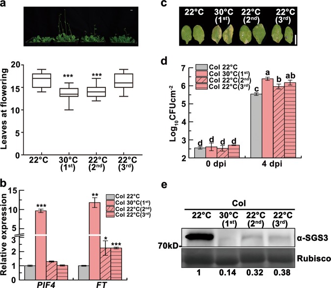
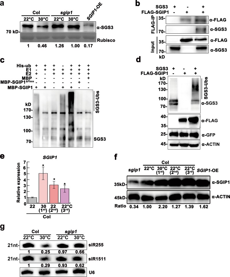
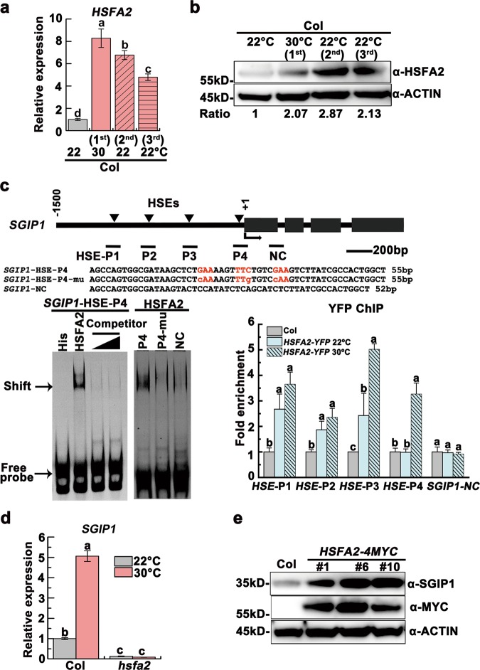
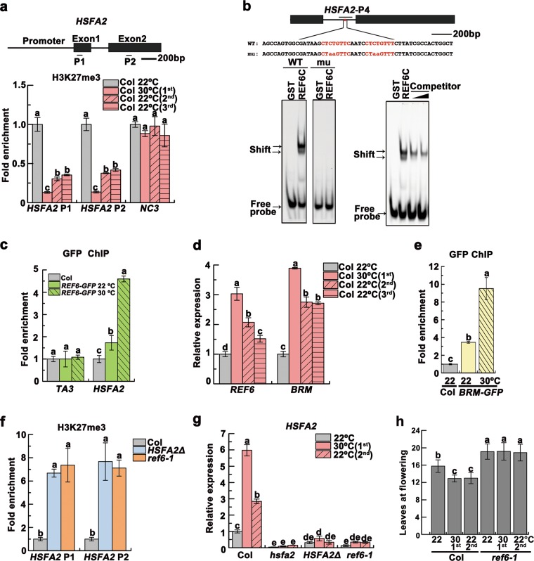
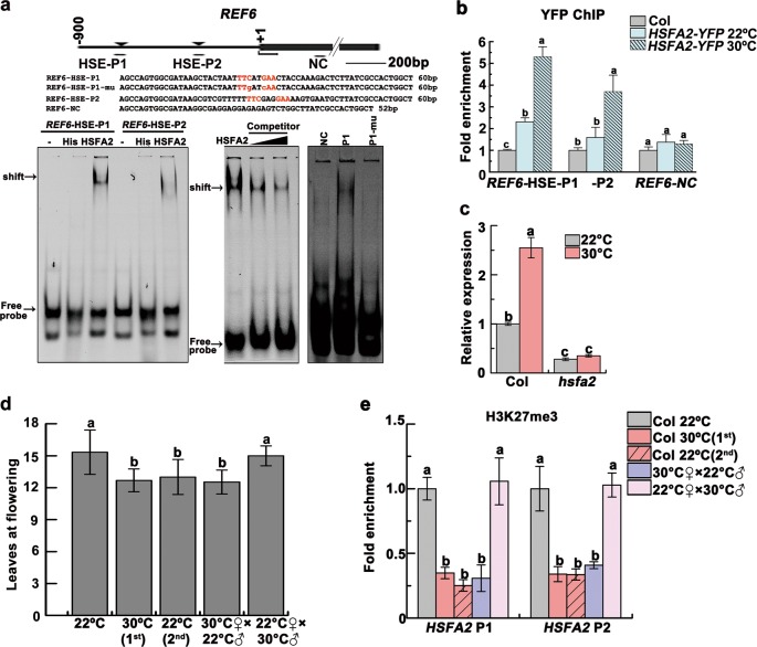
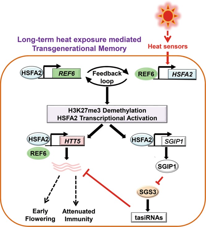
Global warming has profound effects on plant growth and fitness. Plants have evolved sophisticated epigenetic machinery to respond quickly to heat, and exhibit transgenerational memory of the heat-induced release of post-transcriptional gene silencing (PTGS). However, how thermomemory is transmitted to progeny and the physiological relevance are elusive. Here we show that heat-induced HEAT SHOCK TRANSCRIPTION FACTOR A2 (HSFA2) directly activates the H3K27me3 demethylase RELATIVE OF EARLY FLOWERING 6 (REF6), which in turn derepresses HSFA2. REF6 and HSFA2 establish a heritable feedback loop, and activate an E3 ubiquitin ligase, SUPPRESSOR OF GENE SILENCING 3 (SGS3)-INTERACTING PROTEIN 1 (SGIP1). SGIP1-mediated SGS3 degradation leads to inhibited biosynthesis of trans-acting siRNA (tasiRNA). The REF6-HSFA2 loop and reduced tasiRNA converge to release HEAT-INDUCED TAS1 TARGET 5 (HTT5), which drives early flowering but attenuates immunity. Thus, heat induces transmitted phenotypes via a coordinated epigenetic network involving histone demethylases, transcription factors, and tasiRNAs, ensuring reproductive success and transgenerational stress adaptation.


Conflict of interest statement

The authors declare no competing interests.

Figures

Fig. 1
Fig. 1
Heat-induced transgenerational degradation of SGS3 accelerates flowering but attenuates immunity. a Four-week-old 22 °C-grown Col, heat-stressed (first) and unstressed second and third generation plants and box plots of flowering times of these four lines. Flowering time was assessed by counting total leaf numbers in bolting plants (n ≥ 15 for each line). Bars represent means ± SD. b PIF4 and FT transcript levels as normalized to the ACTIN2 signals. The average values (±SD, n = 3) were shown. Samples were collected from 17-day-old seedlings. Asterisks indicate significant difference (Student’s t-test; *P < 0.05, **P < 0.01, ***P < 0.001) (a, b). c Disease symptoms at 4 dpi with Pst DC3000 (avrRpt2). Scale bar, 1 cm (a, c). d Bacterial growth in plants in c was measured at 0 and 4 dpi. Bars represent means ± SD (n = 3). Two-way ANOVA with Tukey’s HSD post hoc test (significance set at P < 0.05) was performed. Different lowercase letters indicate significant differences. e SGS3 levels were immunodetected, with relative levels shown under lanes. Samples were collected from 24-day-old 22 °C-grown Col, heat-stressed (first) and early flowering, unstressed second and third generation plants. Rubisco served as a loading control
Fig. 2
Fig. 2
SGIP1 mediates thermomemory degradation of SGS3 and tasiRNA suppression. a Immunodetection of SGS3 abundance in 24-day-old plants. Coomassie blue staining of Rubisco served as a loading control. b Co-IP assays of SGIP1 with SGS3 in N. benthamiana leaves. Proteins were immunoprecipitated with an anti-FLAG antibody, and detected by western blot using the anti-SGS3 and anti-FLAG antibodies. c In vitro ubiquitination of SGS3 by SGIP1. Polyubiquitination of SGS3 by SGIP1 was detected by immunoblotting using the anti-SGS3 antibody. The mutated SGIP1 protein (SGIP1△Fbox) served as a negative control. d SGIP1 promotes SGS3 polyubiquitination and degradation in vivo. Co-expression of SGIP1 and SGS3 was performed in N. benthamiana leaves. Protein accumulation of SGS3 and FLAG-SGIP1 was immunodetected with the anti-SGS3 and anti-FLAG antibody, respectively. GFP was co-expressed as an internal control. e Expression levels of SGIP1 as normalized to the ACTIN2 signals. Error bars indicate the SD (n = 3). Asterisks indicate significant difference between lines and 22 °C-grown Col (Student’s t-test; *P < 0.05, **P < 0.01). f Immunodetection of SGIP1 levels with an anti-SGIP1 antibody. ACTIN served as a loading control (d, f). g Knockdown of SGIP1 maintained the production of siR255 and siR1511 at 30 °C. U6 was used as a loading control. The intensity of the blots was quantified (a, f, g)
Fig. 3
Fig. 3
HSFA2 activates SGIP1 transgenerationally through binding to its promoter. a, b HSFA2 remained upregulated in the progeny of heat-stressed plants at both transcript (a) and protein (b) levels. c HSFA2 binds to the SGIP1 promoter HSEs in vitro and in vivo. Left: EMSA shows the direct binding of His-HSFA2 to the HSE-P4 of SGIP1. The arrow indicates the shifted bands. Excess unlabeled probe (500 ng and 1000 ng) outcompeted the labeled probe (right lanes). Sequence of the probe is shown and the motif is highlighted in red. A mutant HSE and a downstream sequence SGIP1-NC (negative control) were used as negative controls. Right: ChIP-qPCR validation of HSFA2 occupancy at the HSE regions of the SGIP1 promoter. Data are shown as relative fold enrichments over the background (Col). The SGIP1-NC locus was used as the negative-control locus. The locations of 4 HSEs are indicated by triangles above the gene model. The regions validated by ChIP-qPCR were marked by a bar below the gene model. d SGIP1 transcript levels in Col and hsfa2 plants grown at 22 °C and 30 °C. Gene expression was shown as mean ± SD (n = 3). ACTIN2 was analyzed as an internal control (a, d). Lowercase letters indicate significant differences, as determined by the post hoc Tukey’s HSD test (a, c, d). e The abundance of SGIP1 protein increased in three transgenic lines overexpressing HSFA2-MYC, as immunodetected with anti-SGIP1 and anti-MYC antibodies. ACTIN served as a loading control (b, e) and the signals were quantified (b)
Fig. 4
Fig. 4
H3K27me3 demethylation controls transgenerational upregulation of HSFA2. a H3K27me3 levels detected by ChIP-qPCR at the HSFA2 loci. The gene model was shown and the analyzed regions (P1 and P2) were indicated. One intergenic region (NC3) without H3K27me3 mark was used as negative background. b EMSA showed HSFA2 probe binding to GST-REF6C containing the C2H2-ZnF cluster. The mutant probe abolished the binding of GST-REF6C. Excess unlabeled probe outcompeted the labeled probe (right panel). The motif is highlighted in red in the probe sequence, with mutated bases shown in lowercase. The region validated by ChIP-qPCR in c was marked by a bar. c ChIP-qPCR validation of REF6 binding at HSFA2 using the REF6-GFP plants. Col was used as the negative control. The TA3 locus was used as the negative-control locus. d Transcript levels of REF6 and BRM, shown as mean ± SD (n = 3). e ChIP-qPCR validation of BRM binding at HSFA2 using the BRM-GFP plants. Col was used as the negative control. f H3K27me3 levels of HSFA2 in the HSFA2Δ and ref6–1 plants. The data were normalized to the corresponding input fraction (a, c, e, f). g HSFA2 transcript levels. Data were shown as means ± SD from three replicates (a, c–g). h Flowering times of indicated lines were assessed (n ≥ 15 for each line). Lowercase letters indicate statistical significance based on one-way (a, c–f) or two-way (g, h) ANOVA with Tukey’s HSD post hoc analysis (P < 0.05)
Fig. 5
Fig. 5
REF6 and HSFA2 form a regulatory loop that is maternally transmitted. a EMSA shows that His-HSFA2 protein but not His by itself specifically binds to the first HSE (nTTCnnGAAn motif) of REF6 promoter in vitro. Arrows indicated the shifted bands. Excess unlabeled probe could outcompete the labeled probe (middle). A mutant HSE and a REF6-NC sequence were used as negative controls (right).The motif is highlighted in red in the probe. The region validated by ChIP-qPCR in b was marked by a bar. b ChIP-qPCR validation of HSFA2 occupancy at the REF6 promoter region. The data were normalized to the corresponding input fraction. The REF6-NC locus was used as the negative-control locus. c REF6 transcript level in 24-day-old Col and hsfa2 grown at 22 °C and 30 °C. d The progeny derived from crosses (F1) of Col 30 °C♀×Col 22 °C♂ but not Col 22 °C♀×Col 30 °C♂flowered earlier. Flowering time of different lines was assessed by counting leaf numbers in bolting plants (n ≥ 15 for each line). e Lower H3K27me3 levels of HSFA2 persisted in the progeny derived from Col 30 °C♀×Col 22 °C♂but not Col 22 °C♀×Col 30 °C♂. Data were shown as means ± SD from three replicates (b, c, e). Lowercase letters indicate statistical significance based on one-way (b, d, e) or two-way (c) ANOVA with Tukey’s HSD post hoc analysis (P < 0.05)
Fig. 6
Fig. 6
HTT5 coordinates thermomemory phenotypes via reduced tasiRNAs and the REF6-HSFA2 loop. a ChIP-qPCR validation of HSFA2 (left), REF6 (middle) and BRM (right) occupancy at the HTT5 loci. The analyzed regions were marked by bars. b HTT5 transcript levels in 24-day-old Col, hsfa2, ref6–1, brm-1 and ref6–1brm-1 grown at 22 °C and 30 °C. c Heat-induced upregulation of HTT5 could be transmitted to progeny, as detected by qRT-PCR. d Flowering times of indicated lines were assessed (n ≥ 15 for each line). e Disease symptoms at 4 dpi with Pst DC3000 (avrRpt2). Scale bar, 1 cm. f Bacterial growth was measured. Data are shown as means ± SD from three replicates (a, b, c, f). Lowercase letters indicate statistical significance based on one-way (a, c, d) or two-way (b, f) ANOVA with Tukey’s HSD post hoc analysis (P < 0.05)
Fig. 7
Fig. 7
A proposed model depicting transgenerational thermomemory that is mediated by the H3K27me3 demethylation-HSFA2 regulatory loop in Arabidopsis. Long-term heat exposure activates plant heat sensor(s) that upregulates HSFA2, which targets and activates H3K27me3 demethylase REF6 and the chromatin remodeler BRM. REF6/BRM in turn triggers H3K27me3 demethylation to activate HSFA2. Thus, REF6 and HSFA2 form a heritable transcriptional feedback loop in heat responses and memory. This regulatory loop activates the E3 ligase, SGIP1, to trigger the transgenerational degradation of SGS3, leading to the suppression of tasiRNA biosynthesis. The REF6-HSFA2 loop and reduced tasiRNA levels converge to activate HTT5, which coordinates early flowering with decreased disease resistance in heat-stressed plants and progeny

References

    1. Pauli H, et al. Recent plant diversity changes on Europe’s mountain summits. Science. 2012;336:353–355. doi: 10.1126/science.1219033. - DOI - PubMed
    1. Lobell DB, Schlenker W, Costa-Roberts J. Climate trends and global crop production since 1980. Science. 2011;333:616–620. doi: 10.1126/science.1204531. - DOI - PubMed
    1. Liu J, Feng L, Li J, He Z. Genetic and epigenetic control of plant heat responses. Front. Plant Sci. 2015;6:267. - PMC - PubMed
    1. Tittel-Elmer M, et al. Stress-induced activation of heterochromatic transcription. PLoS Genet. 2010;6:e1001175. doi: 10.1371/journal.pgen.1001175. - DOI - PMC - PubMed
    1. Pecinka A, et al. Epigenetic regulation of repetitive elements is attenuated by prolonged heat stress in Arabidopsis. Plant Cell. 2010;22:3118–3129. doi: 10.1105/tpc.110.078493. - DOI - PMC - PubMed

Publication types

MeSH terms