. 2019 Feb 4:13:11.
doi: 10.3389/fncel.2019.00011. eCollection 2019.

A Conditioning Sciatic Nerve Lesion Triggers a Pro-regenerative State in Primary Sensory Neurons Also of Dorsal Root Ganglia Non-associated With the Damaged Nerve

Affiliations

A Conditioning Sciatic Nerve Lesion Triggers a Pro-regenerative State in Primary Sensory Neurons Also of Dorsal Root Ganglia Non-associated With the Damaged Nerve

Petr Dubový et al. Front Cell Neurosci. .

Abstract

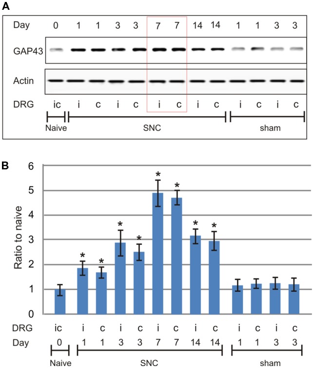
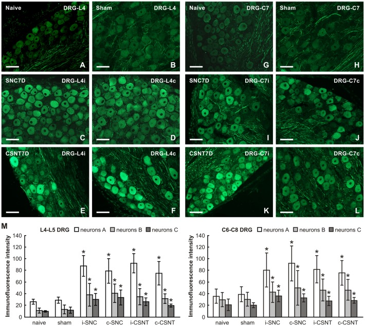
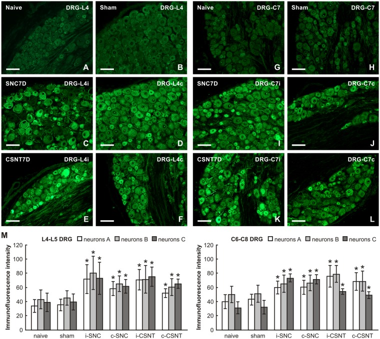
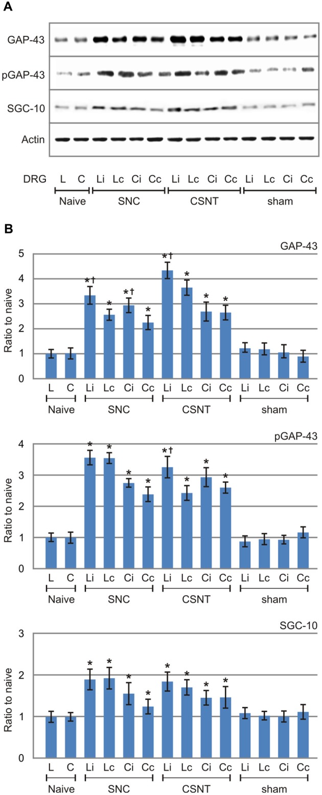
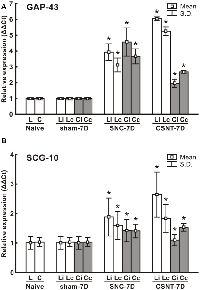
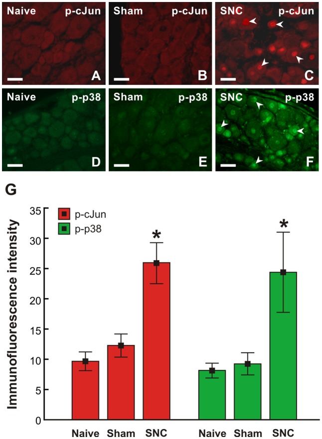
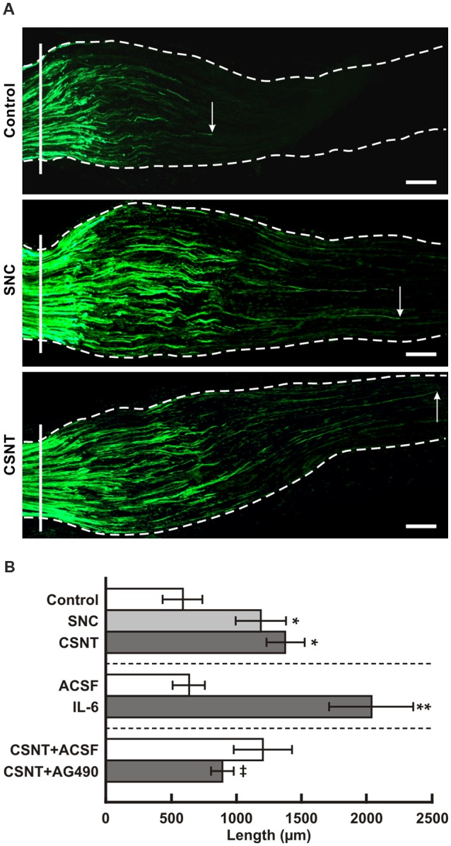
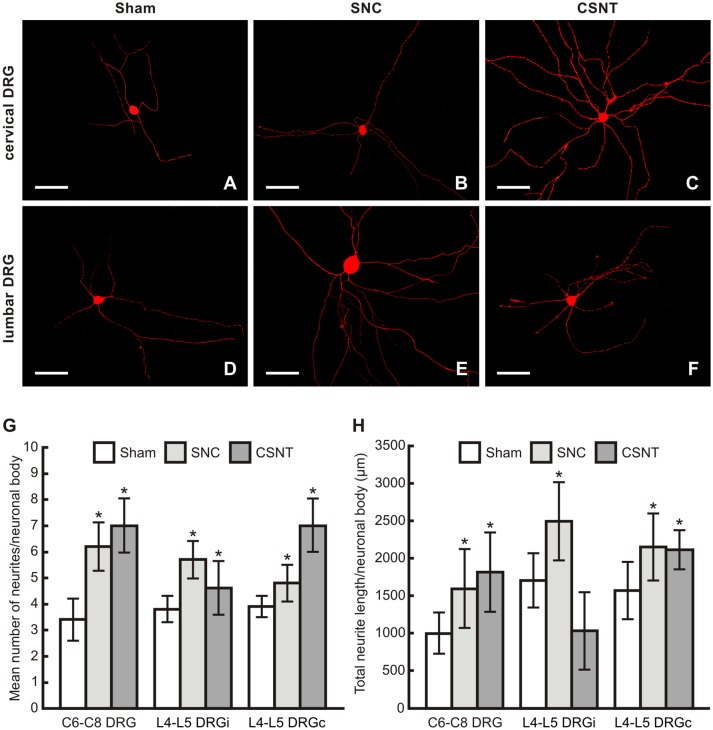
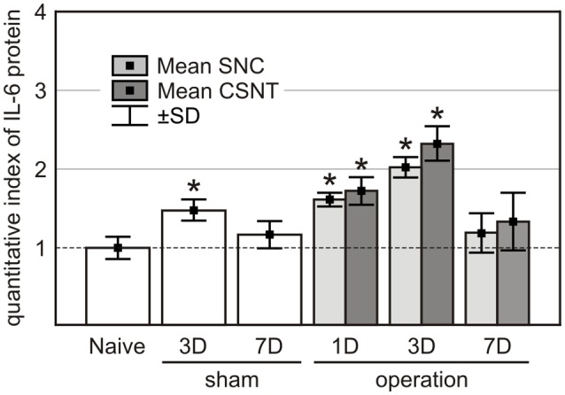
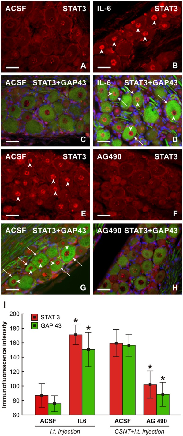
The primary sensory neurons of dorsal root ganglia (DRG) are a very useful model to study the neuronal regenerative program that is a prerequisite for successful axon regeneration after peripheral nerve injury. Seven days after a unilateral sciatic nerve injury by compression or transection, we detected a bilateral increase in growth-associated protein-43 (GAP-43) and superior cervical ganglion-10 (SCG-10) mRNA and protein levels not only in DRG neurons of lumbar spinal cord segments (L4-L5) associated with injured nerve, but also in remote cervical segments (C6-C8). The increase in regeneration-associated proteins in the cervical DRG neurons was associated with the greater length of regenerated axons 1 day after ulnar nerve crush following prior sciatic nerve injury as compared to controls with only ulnar nerve crush. The increased axonal regeneration capacity of cervical DRG neurons after a prior conditioning sciatic nerve lesion was confirmed by neurite outgrowth assay of in vitro cultivated DRG neurons. Intrathecal injection of IL-6 or a JAK2 inhibitor (AG490) revealed a role for the IL-6 signaling pathway in activating the pro-regenerative state in remote DRG neurons. Our results suggest that the pro-regenerative state induced in the DRG neurons non-associated with the injured nerve reflects a systemic reaction of these neurons to unilateral sciatic nerve injury.

Keywords: GAP-43; IL-6; SCG-10; neurite outgrowth assay; primary sensory neurons; pro-regenerative state; ulnar nerve crush; unilateral nerve injury.


Figures

Figure 1
Figure 1
Results of western blot analysis of growth-associated protein-43 (GAP-43) protein levels in cervical dorsal root ganglia (DRG; C6-C8) removed from naïve rats as well as sham- and sciatic nerve compression (SNC)-operated rats. Sham-operated rats were left to survive for 1 and 3 days, SNC-operated rats for 1, 3, 7 and 14 days (n = 3 for each group). Upper panel (A) shows a representative western blot with GAP-43 protein in cervical DRG ipsilateral (i) and contralateral (c) to unilateral SNC. Equal loading of proteins was confirmed by actin levels (Actin). Lower panel (B) shows densitometry of the individual protein bands after normalization to actin; the intensities of the bands from naïve cervical DRG were as arbitrarily set to 1. *Significant difference (p < 0.05) compared to naïve or sham-operated rats in a Mann-Whitney U-test.
Figure 2
Figure 2
Representative pictures of cryostat sections through the DRG from naïve (A,G), sham-operated (B,H) rats and rats with unilateral sciatic nerve compression (SNC; C,D,I,J) or complete sciatic nerve transection (CSNT; E,F,K,L) for 7 days. The sections of ipsilateral (C,E) and contralateral (D,F) DRG of the L4 spinal segment as well as ipsilateral (I,K) and contralateral (J,L) DRG of the C7 spinal segment were incubated under the same conditions with mouse monoclonal antibody recognizing GAP-43. Scale bars = 75 μm. (M) Graph illustrating the mean intensity of GAP-43 immunofluorescence in individual DRG neuron-size classes of cervical (C6-C8) and lumbar (L4-L5) spinal segments ipsilateral (i) and contralateral (c) to unilateral SNC and transection (CSNT) for 7 days; n = 6 for each group. Neurons A ≥ 40 μm; Neurons B 25–40 μm; Neurons C ≤ 25 μm. *Significant difference (p < 0.05) compared to naive or sham-operated rats in a Mann-Whitney U-test.
Figure 3
Figure 3
Representative pictures of cryostat sections through the DRG from naïve (A,G), sham-operated (B,H) rats and rats with unilateral SNC (C,D,I,J) or CSNT (E,F,K,L) for 7 days. The sections of ipsilateral (C,E) and contralateral (D,F) DRG of the L4 spinal segment as well as ipsilateral (I,K) and contralateral (J,L) DRG of the C7 spinal segment were incubated under the same conditions with rabbit polyclonal antibody recognizing superior cervical ganglion-10 (SCG-10). Scale bars = 75 μm. (M) Graph illustrating the mean intensity of SCG-10 immunofluorescence measured in individual DRG neuron-size classes of cervical (C6-C8) and lumbar (L4-L5) spinal segments ipsilateral (i) and contralateral (c) to unilateral SNC and transection (CSNT) for 7 days; n = 6 for each group. Neurons A ≥ 40 μm; Neurons B 25–40 μm; Neurons C ≤ 25 μm. *Significant difference (p < 0.05) compared to naive or sham-operated rats in a Mann-Whitney U-test.
Figure 4
Figure 4
Results of western blot analysis of GAP-43, pGAP-43 and SCG-10 protein levels in DRG of L4-L5 (L) and C6-C8 (C) segments removed from ipsilateral (i) and contralateral (c) sides of naïve as well as sham-, SNC- and CSNT-operated rats for 7 days. Upper panel (A) illustrates representative western blots of DRG from three rats for each group. Equal loading of proteins was confirmed by actin levels (Actin). The same Actin controls were used for analysis of GAP-43, pGAP-43 and SCG-10 protein levels in this set representative western blots. Lower panels (B) show densitometry of individual protein bands after normalization to actin from three independent experiments; the intensities of the bands from naïve DRG were as arbitrarily set to 1. *Significant difference (p < 0.05) when compared to sham-operated rats; Significant difference (p < 0.05) compared to contralateral DRG in a Mann-Whitney U-test.
Figure 5
Figure 5
Results of real-time PCR (RT-PCR) of relative GAP-43 (A) and SCG-10 (B) mRNA levels in DRG of lumbar (L4-L5) and cervical (C6-C8) spinal segments of removed from ipsilateral (i) and contralateral (c) sides of Naïve as well as sham-, SNC- and CSNT-operated rats for 7 days; n = 6 for each group. Relative expressions were determined using Actin as the housekeeping gene and normalized to naïve controls. *Significant difference (p < 0.05) compared to sham-operated rats in a Mann-Whitney U-test.
Figure 6
Figure 6
Representative pictures of cervical (C7) DRG neurons of intact rats (A–D) and rats 7 day after sham- (B,E) or SNC-operation (C,F). DRG sections were immunostained for p-cJun (A–C) and p-p38 (D–F). Scale bars = 40 μm. (G) Graph illustrating mean immunofluorescence intensity of activated p-cJun (red columns) and p-p38 (green columns) in cervical DRG neurons of naïve, sham, and SNC groups. SNC induced activation of p-cJun and p-p38 (arrowheads, C,G). *Significant difference (p < 0.05) compared to naïve or sham-operated rats in a Mann-Whitney U-test.
Figure 7
Figure 7
(A) Representative longitudinal sections through the ulnar nerves distal to the crush site (solid line) showing SCG-10 positive regenerated axons. Arrows indicate the tip of the longest SCG-10 positive axons. The ulnar nerves were removed after 1 day from control rat (only ulnar nerve crush) and from rats 7 days after prior SNC or CSNT. Scale bars = 100 μm. (B) The top portion of the graph illustrates mean length of regenerated SCG10+ axons ± SD in the ulnar nerve 1 day after crush in rats without a sciatic nerve injury (Control) and following 7 days from SNC or CSNT; n = 6 for each group. *Significant difference (p < 0.05) compared to control. The middle portion illustrates mean length of regenerated SCG10+ axons ± SD in the ulnar nerve 1 day after crush and intrathecal injection of 10 μl of artificial cerebrospinal fluid (ACSF) or IL-6 (20 ng/10 μl); n = 4 for each group. **Significant difference (p < 0.05) compared to control. The bottom portion illustrates mean length of regenerated SCG10+ axons ± SD in the ulnar nerve 1 day after crush 7 days from prior CSNT and intrathecal application of ACSF (CSNT+ACSF) or JAK2 inhibitor (CSNT+AG490); n = 4 for each group. Significant difference (p < 0.05) compared to CSNT+ACSF group in a Mann-Whitney U-test.
Figure 8
Figure 8
(A–F) Representative pictures of cervical (C6-C8) and lumbar (L4-L5) DRG neurons of the ipsilateral side dissociated and cultured in vitro after removing of DRG from sham-, SNC- or CSNT-operated rats for 7 days. The DRG neurons were fixed and immunostained for βIII-tubulin after 2 days of in vitro incubation. Scale bars = 75 μm. The graphs illustrate mean number of neurites (G) and total neurite length (H) per neuronal body of in vitro cultivated DRG neurons of cervical (C6-C8) and lumbar (L4-L5) spinal segments from the ipsilateral (i) and contralateral (c) sides removed from sham-, SNC- and CSNT-operated rats (n = 4 for each group). At least 100 randomly selected neurons with nuclei per cover slip were measured. *Significant difference (p < 0.05) compared to sham-operated controls in a Mann-Whitney U-test.
Figure 9
Figure 9
IL-6 protein levels in the plasma of naïve, sham-, SNC- and CSNT-operated rats (n = 3 for each group). Blood samples of sham-operated rats were obtained 3 (3D) and 7 (7D) days after surgical treatment and those from SNC- and CSNT-operated rats were obtained 1 (1D), 3 (3D), and 7 (7D) days after operation. *Significant difference (P < 0.05) compared to baseline levels of naive rats in a Mann-Whitney U-test.
Figure 10
Figure 10
Representative pictures of cervical (C7) DRG neurons of intact rats (A–D) and rats 1 day after the ulnar nerve crush following 7 days from CSNT (E–H). In intact rats, 10 μl of ACSF (A,C) or IL-6 (B,D) were intrathecally applied for 1 day. Cervical DRG were also removed from rats with CSNT for 7 days, a subsequent ulnar nerve crush for 1 day and intrathecal injection of 10 μl of ACSF (E,G) or AG490 (F,H). DRG sections were immunostained for STAT3 (A,B,E,F) or double immunostained (C,D,G,H) for STAT3 (red fluorescence) and GAP-43 (green fluorescence). Intrathecal injection of IL-6 induced activation and nuclear translocation of STAT3 (arrowheads, B,D) as well as increased immunostaining for GAP-43 in neurons (arrows, D) when compared with ACSF treatment (C). Cervical DRG neurons of rats following the ulnar nerve crush 7 days after CSNT displayed activation and nuclear translocation of STAT3 (E,G) and intense GAP-43 immunofluorescence in the neurons when injected with ACSF (G, arrowheads and arrows, respectively) while intrathecal application of AG490 resulted in a marked reduction of STAT3 activation and GAP-43 immunostaining (F,H). Scale bars = 40 μm. (I) Graph illustrating mean immunofluorescence intensity of activated STAT3 and GAP-43 in cervical DRG neurons of intact rats following intrathecal application of ACSF or IL-6 for 1 day and rats with CSNT for 7 days and intrathecal injection of ACSF or AG490. *Significant difference (P < 0.05) compared with ACSF-treated rats in a Mann-Whitney U-test.

References

    1. Abe N., Borson S. H., Gambello M. J., Wang F., Cavalli V. (2010). Mammalian target of rapamycin (mTOR) activation increases axonal growth capacity of injured peripheral nerves. J. Biol. Chem. 285, 28034–28043. 10.1074/jbc.m110.125336 - DOI - PMC - PubMed
    1. Al-Ali H., Beckerman S. R., Bixby J. L., Lemmon V. P. (2017). In vitro models of axon regeneration. Exp. Neurol. 287, 423–434. 10.1016/j.expneurol.2016.01.020 - DOI - PMC - PubMed
    1. Bareyre F. M., Garzorz N., Lang C., Misgeld T., Buning H., Kerschensteiner M. (2011). In vivo imaging reveals a phase-specific role of STAT3 during central and peripheral nervous system axon regeneration. Proc. Natl. Acad. Sci. U S A 108, 6282–6287. 10.1073/pnas.1015239108 - DOI - PMC - PubMed
    1. Bonilla I. E., Tanabe K., Strittmatter S. M. (2002). Small proline-rich repeat protein 1A is expressed by axotomized neurons and promotes axonal outgrowth. J. Neurosci. 22, 1303–1315. 10.1523/jneurosci.22-04-01303.2002 - DOI - PMC - PubMed
    1. Brázda V., Klusáková I., Svíženská I. H., Dubovy P. (2013). Dynamic response to peripheral nerve injury detected by in situ hybridization of IL-6 and its receptor mRNAs in the dorsal root ganglia is not strictly correlated with signs of neuropathic pain. Mol. Pain 9:42. 10.1186/1744-8069-9-42 - DOI - PMC - PubMed

LinkOut - more resources