. 2019 Apr;97(4):533-540.
doi: 10.1007/s00109-019-01755-3. Epub 2019 Feb 18.

Molecular modeling of LDLR aids interpretation of genomic variants

Affiliations

Molecular modeling of LDLR aids interpretation of genomic variants

Eric W Klee et al. J Mol Med (Berl). 2019 Apr.

Abstract

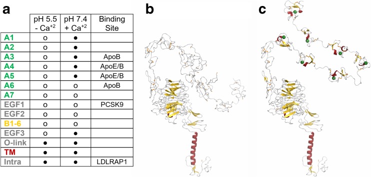
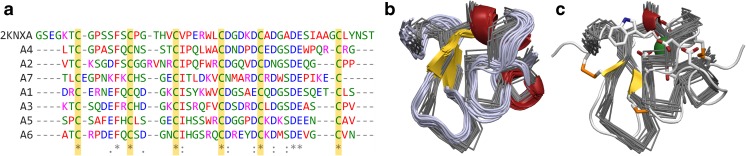
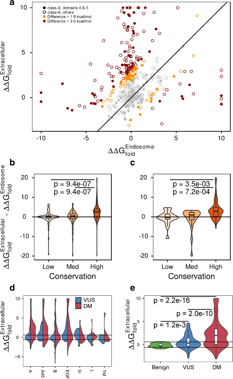
Genetic variants in low-density lipoprotein receptor (LDLR) are known to cause familial hypercholesterolemia (FH), occurring in up to 1 in 200 people (Youngblom E. et al. 1993 and Nordestgaard BG et al. 34:3478-3490a, 2013) and leading to significant risk for heart disease. Clinical genomics testing using high-throughput sequencing is identifying novel genomic variants of uncertain significance (VUS) in individuals suspected of having FH, but for whom the causal link to the disease remains to be established (Nordestgaard BG et al. 34:3478-3490a, 2013). Unfortunately, experimental data about the atomic structure of the LDL binding domains of LDLR at extracellular pH does not exist. This leads to an inability to apply protein structure-based methods for assessing novel variants identified through genetic testing. Thus, the ambiguities in interpretation of LDLR variants are a barrier to achieving the expected clinical value for personalized genomics assays for management of FH. In this study, we integrated data from the literature and related cellular receptors to develop high-resolution models of full-length LDLR at extracellular conditions and use them to predict which VUS alter LDL binding. We believe that the functional effects of LDLR variants can be resolved using a combination of structural bioinformatics and functional assays, leading to a better correlation with clinical presentation. We have completed modeling of LDLR in two major physiologic conditions, generating detailed hypotheses for how each of the 1007 reported protein variants may affect function. KEY MESSAGES: • Hundreds of variants are observed in the LDLR, but most lack interpretation. • Molecular modeling is aided by biochemical knowledge. • We generated context-specific 3D protein models of LDLR. • Our models allowed mechanistic interpretation of many variants. • We interpreted both rare and common genomic variants in their physiologic context. • Effects of genomic variants are often context-specific.

Keywords: Familial hypercholesterolemia; Genomic interpretation; Low-density lipoprotein receptor; Molecular modeling; Variant prioritization.


Figures

Fig. 1
Fig. 1
Structural model of LDLR at extracellular conditions generated by date integration and molecular modeling. A Each LDLR domain is available (o) or modeled by us (●). We have used molecular modeling to generate full-length LDLR structures at B endosomal and C extracellular conditions by leveraging available experimental data
Fig. 2
Fig. 2
Conservation and modeling of LDLR Class-A domains. A The sequence alignment of class-A domains with the sequence of LRP-1 class-A domain, colored by amino acid type, shows the conservation of six cysteine residues and a pattern of acidic residues. These six cysteine residues form three disulfide bonds and the acidic residues form a binding pocket for Ca+2. B The backbone of LRP-1 class-A domain, solved by NMR, is shown in gray. All models from the NMR ensemble are shown. Superimposed and shown in cartoon representation, colored by secondary structure type, is LRP-1 class-A domain bound to the minimal peptide from ApoB. The overall fold is strikingly similar between bound and unbound conformations. C The backbone ribbons of LRP-1 are shown again, but now superimposed onto the fourth class-A domain of our extracellular LDLR model. The three disulfide bonds are shown in orange, Ca+2 green, and the residues interacting with the Ca+2 ion are shown in detail
Fig. 3
Fig. 3
LDLR variants have context-specific effects. Each variant may confer significantly different effects on protein stability between endosomal and extracellular conditions. A Each data point represents a different LDLR variant. We evaluated 403 unique missense genomic variants observed in population (ExAC) or disease (ClinVar or HGMD) databases within the class-A domains. Symbols are filled in for the 128 variants from the fourth and fifth class-A domains. The line of equivalence is shown and variants colored gray if they exhibit a difference of less than 1.8 kcal/mol. The 57 (14%) of variants with a difference between 1.8 and 3.0 kcal/mol are colored orange, and the 119 (30%) variants with a difference greater than 3.0 kcal/mol colored red. B Across all class-A domains, there is a significant relationship between residue conservation and the difference in stability between conditions. C This relationship is present within the fourth and fifth class-A domains. D Across LDLR domains, missense variants in the class-A domains have the strongest separation in ΔΔGfold between pathogenic variants and VUS. Horizontal lines mark 0.6 kcal/mol. Pathogenic missense variants in all extracellular domains are more likely to be destabilizing to the native structure compared to VUS. Many VUS in the fourth and fifth class-A and EGF domains are destabilizing. E For our extracellular model of class-A domains, there are strong differences between the distribution of ΔΔGfold among benign, VUS, and pathogenic variants. Not all pathogenic variants destabilize the conformation, but a significant fraction does. A smaller, but still significant proportion of VUS is destabilizing, but no benign variants are destabilizing

References

    1. Youngblom E, Pariani M, Knowles JW (1993) Familial hypercholesterolemia. In: Adam MP, Ardinger HH, Pagon RA et al. (eds) GeneReviews((R)). Seattle (WA),
    1. Nordestgaard BG, Chapman MJ, Humphries SE, Ginsberg HN, Masana L, Descamps OS, Wiklund O, Hegele RA, Raal FJ, Defesche JC, Wiegman A, Santos RD, Watts GF, Parhofer KG, Hovingh GK, Kovanen PT, Boileau C, Averna M, Boren J, Bruckert E, Catapano AL, Kuivenhoven JA, Pajukanta P, Ray K, Stalenhoef AF, Stroes E, Taskinen MR, Tybjaerg-Hansen A, European Atherosclerosis Society Consensus P Familial hypercholesterolaemia is underdiagnosed and undertreated in the general population: guidance for clinicians to prevent coronary heart disease: consensus statement of the European Atherosclerosis Society. Eur Heart J. 2013;34(45):3478–3490a. doi: 10.1093/eurheartj/eht273. - DOI - PMC - PubMed
    1. Fairoozy RH, Futema M, Vakili R, Abbaszadegan MR, Hosseini S, Aminzadeh M, Zaeri H, Mobini M, Humphries SE, Sahebkar A. The genetic spectrum of familial hypercholesterolemia (FH) in the Iranian population. Sci Rep. 2017;7(1):17087. doi: 10.1038/s41598-017-17181-9. - DOI - PMC - PubMed
    1. Martinez-Olivan J, Arias-Moreno X, Velazquez-Campoy A, Millet O, Sancho J. LDL receptor/lipoprotein recognition: endosomal weakening of ApoB and ApoE binding to the convex face of the LR5 repeat. FEBS J. 2014;281(6):1534–1546. doi: 10.1111/febs.12721. - DOI - PubMed
    1. Fass D, Blacklow S, Kim PS, Berger JM. Molecular basis of familial hypercholesterolaemia from structure of LDL receptor module. Nature. 1997;388(6643):691–693. doi: 10.1038/41798. - DOI - PubMed

LinkOut - more resources