

White matter tracts lesions and decline of verbal fluency after deep brain stimulation in Parkinson's disease
- PMID: 30779251
- PMCID: PMC6865750
- DOI: 10.1002/hbm.24544
White matter tracts lesions and decline of verbal fluency after deep brain stimulation in Parkinson's disease
Abstract
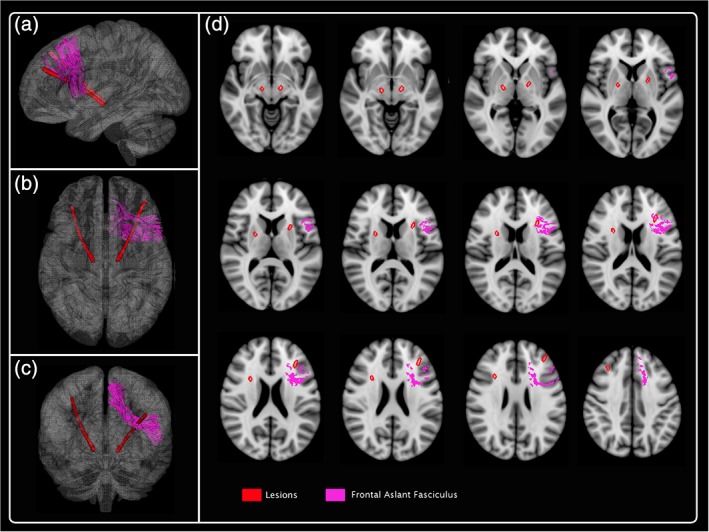
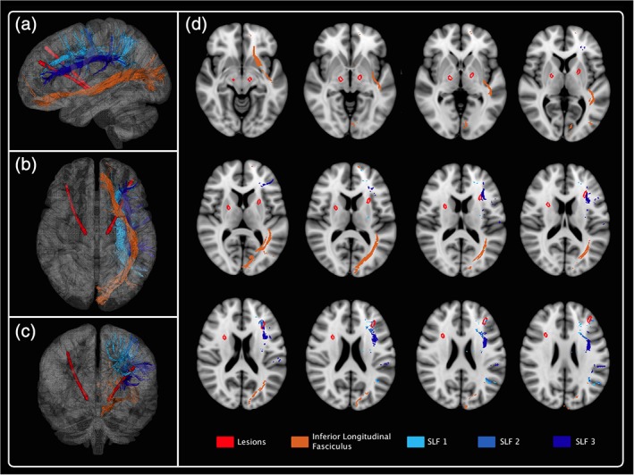
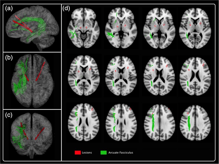
Decline of verbal fluency (VF) performance is one of the most systematically reported neuropsychological adverse effects after subthalamic nucleus deep brain stimulation (STN-DBS). It has been suggested that this worsening of VF may be related to a microlesion due to the electrode trajectories. We describe the disruption of surrounding white matter tracts following electrode implantation in Parkinson's disease (PD) patients with STN-DBS and assess whether damage of fiber pathways is associated with VF impairment after surgery. We retrospectively analyzed 48 PD patients undergoing bilateral STN DBS. The lesion mask along the electrode trajectory transformed into the MNI 152 coordinate system, was compared with white matter tract atlas in Tractotron software, which provides a probability and proportion of fibers disconnection. Combining tract- and atlas-based analysis reveals that the trajectory of the electrodes intersected successively with the frontal aslant tract, anterior segment of arcuate tract, the long segment of arcuate tract, the inferior longitudinal fasciculus, the superior longitudinal fasciculus, the anterior thalamic radiation, and the fronto striatal tract. We found no association between the proportion fiber disconnection and the severity of VF impairment 6 months after surgery. Our findings demonstrated that microstructural injury associated with electrode trajectories involved white matter bundles implicated in VF networks.
Keywords: Parkinson's disease; deep brain stimulation; microlesion; subthalamic nucleus; verbal fluency; white matter tracts.
© 2019 Wiley Periodicals, Inc.
Conflict of interest statement
The authors declare that they have no conflict of interest.
Figures

Similar articles
-
Decline in verbal fluency after subthalamic nucleus deep brain stimulation in Parkinson's disease: a microlesion effect of the electrode trajectory?J Parkinsons Dis. 2015;5(1):95-104. doi: 10.3233/JPD-140443. J Parkinsons Dis. 2015. PMID: 25374271
-
Anterior lead location predicts verbal fluency decline following STN-DBS in Parkinson's disease.Parkinsonism Relat Disord. 2021 Nov;92:36-40. doi: 10.1016/j.parkreldis.2021.10.012. Epub 2021 Oct 14. Parkinsonism Relat Disord. 2021. PMID: 34678718
-
The impact of white matter lesions on the cognitive outcome of subthalamic nucleus deep brain stimulation in Parkinson's disease.Clin Neurol Neurosurg. 2017 Aug;159:87-92. doi: 10.1016/j.clineuro.2017.05.023. Epub 2017 May 31. Clin Neurol Neurosurg. 2017. PMID: 28582689
-
Cognition and Depression Following Deep Brain Stimulation of the Subthalamic Nucleus and Globus Pallidus Pars Internus in Parkinson's Disease: A Meta-Analysis.Neuropsychol Rev. 2015 Dec;25(4):439-54. doi: 10.1007/s11065-015-9302-0. Epub 2015 Oct 12. Neuropsychol Rev. 2015. PMID: 26459361 Review.
-
Congress of Neurological Surgeons Systematic Review and Evidence-Based Guideline on Subthalamic Nucleus and Globus Pallidus Internus Deep Brain Stimulation for the Treatment of Patients With Parkinson's Disease: Executive Summary.Neurosurgery. 2018 Jun 1;82(6):753-756. doi: 10.1093/neuros/nyy037. Neurosurgery. 2018. PMID: 29538685 Free PMC article.
Cited by
-
Effects of Deep Brain Stimulation in the Subthalamic Nucleus on Neurocognitive Function in Patients With Parkinson's Disease Compared With Medical Therapy: A Meta-Analysis.Front Neurol. 2021 Mar 2;12:610840. doi: 10.3389/fneur.2021.610840. eCollection 2021. Front Neurol. 2021. PMID: 33737902 Free PMC article.
-
Neuropsychiatric Symptoms in Parkinson's Disease After Subthalamic Nucleus Deep Brain Stimulation.Front Neurol. 2021 May 4;12:656041. doi: 10.3389/fneur.2021.656041. eCollection 2021. Front Neurol. 2021. PMID: 34017303 Free PMC article.
-
Differential cognitive effects of unilateral left and right subthalamic nucleus deep brain stimulation for Parkinson disease.medRxiv [Preprint]. 2023 Mar 1:2023.02.27.23286478. doi: 10.1101/2023.02.27.23286478. medRxiv. 2023. Update in: Ann Neurol. 2024 Jun;95(6):1205-1219. doi: 10.1002/ana.26903. PMID: 36909562 Free PMC article. Updated. Preprint.
-
Altered Spontaneous Neural Activity and Functional Connectivity in Parkinson's Disease With Subthalamic Microlesion.Front Neurosci. 2021 Jul 20;15:699010. doi: 10.3389/fnins.2021.699010. eCollection 2021. Front Neurosci. 2021. PMID: 34354566 Free PMC article.
-
A narrative review of the anatomy and function of the white matter tracts in language production and comprehension.Front Hum Neurosci. 2023 Mar 27;17:1139292. doi: 10.3389/fnhum.2023.1139292. eCollection 2023. Front Hum Neurosci. 2023. PMID: 37051488 Free PMC article. Review.
References
-
- Biesbroek, J. M. , van Zandvoort, M. J. , Kappelle, L. J. , Velthuis, B. K. , Biessels, G. J. , & Postma, A. (2016). Shared and distinct anatomical correlates of semantic and phonemic fluency revealed by lesion‐symptom mapping in patients with ischemic stroke. Brain Structure and Function, 221(4), 2123–2134. 10.1007/s00429-015-1033-8 - DOI - PMC - PubMed
-
- Birn, R. M. , Kenworthy, L. , Case, L. , Caravella, R. , Jones, T. B. , Bandettini, P. A. , & Martin, A. (2010). Neural systems supporting lexical search guided by letter and semantic category cues: A self‐paced overt response fMRI study of verbal fluency. Neuroimage, 49(1), 1099–1107. 10.1016/j.neuroimage.2009.07.036 - DOI - PMC - PubMed
MeSH terms
LinkOut - more resources
Full Text Sources
Medical
