

Molecular characterization and expression of sensory neuron membrane proteins in the parasitoid Microplitis mediator (Hymenoptera: Braconidae)
- PMID: 30779304
- PMCID: PMC7277062
- DOI: 10.1111/1744-7917.12667
Molecular characterization and expression of sensory neuron membrane proteins in the parasitoid Microplitis mediator (Hymenoptera: Braconidae)
Abstract
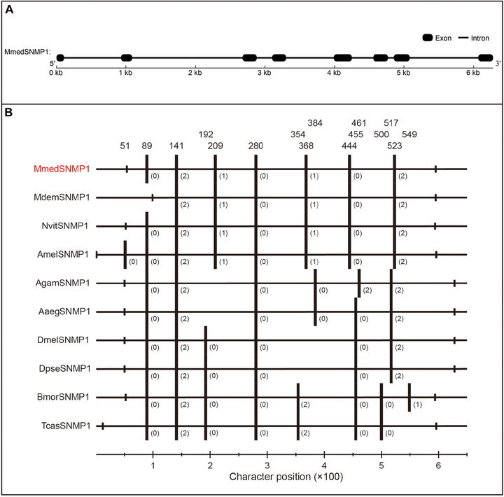
Sensory neuron membrane proteins (SNMPs), homologs of the human fatty acid transport protein CD36 family, are observed to play a significant role in chemoreception, especially in detecting sex pheromone in Drosophila and some lepidopteran species. In the current study, two full-length SNMP transcripts, MmedSNMP1 and MmedSNMP2, were identified in the parasitoid Microplitis mediator (Hymenoptera: Braconidae). Quantitative real-time polymerase chain reaction analysis showed that the expression of MmedSNMP1 was significantly higher in antennae than in other tissues of both sexes. In addition, the MmedSNMP1 transcript was increased dramatically in newly emerged adults and there were no significant differences between adults with or without mating and parasitic experiences. However, compared with MmedSNMP1, the expression of MmedSNMP2 was widely found in various tissues, significantly increased at half-pigmented pupae stage and remained at a relatively constant level during the following developmental stages. It was found that MmedSNMP1 contained eight exons and seven introns, which was highly conserved compared with other insect species. In situ hybridization assay demonstrated that MmedSNMP1 transcript was distributed widely in antennal flagella. Among selected chemosensory genes (odorant binding protein, odorant receptor, and ionotropic receptor genes), MmedSNMP1 only partially overlapped with MmedORco in olfactory sensory neurons of antennae. Subsequent immunolocalization results further indicated that MmedSNMP1 was mainly expressed in sensilla placodea of antennae and possibly involved in perceiving plant volatiles and sex pheromones. These findings lay a foundation for further investigating the roles of SNMPs in the chemosensation of parasitoids.
Keywords: Microplitis mediator; antennae; chemosensation; expression profile; in situ hybridization; sensory neuron membrane proteins.
© 2019 Institute of Zoology, Chinese Academy of Sciences.
Figures


Similar articles
-
Identification and Expression Analysis of Putative Chemosensory Receptor Genes in Microplitis mediator by Antennal Transcriptome Screening.Int J Biol Sci. 2015 May 16;11(7):737-51. doi: 10.7150/ijbs.11786. eCollection 2015. Int J Biol Sci. 2015. PMID: 26078716 Free PMC article.
-
Characterization of antennal chemosensilla and associated odorant binding as well as chemosensory proteins in the parasitoid wasp Microplitis mediator (Hymenoptera: Braconidae).Sci Rep. 2018 May 16;8(1):7649. doi: 10.1038/s41598-018-25996-3. Sci Rep. 2018. PMID: 29769575 Free PMC article.
-
Molecular identification and differential expression of sensory neuron membrane proteins in the antennae of the black cutworm moth Agrotis ipsilon.J Insect Physiol. 2013 Apr;59(4):430-43. doi: 10.1016/j.jinsphys.2013.02.003. Epub 2013 Feb 21. J Insect Physiol. 2013. PMID: 23454276
-
The role of SNMPs in insect olfaction.Cell Tissue Res. 2021 Jan;383(1):21-33. doi: 10.1007/s00441-020-03336-0. Epub 2020 Nov 27. Cell Tissue Res. 2021. PMID: 33245414 Free PMC article. Review.
-
Comprehensive History of CSP Genes: Evolution, Phylogenetic Distribution and Functions.Genes (Basel). 2020 Apr 10;11(4):413. doi: 10.3390/genes11040413. Genes (Basel). 2020. PMID: 32290210 Free PMC article. Review.
Cited by
-
Silencing sensory neuron membrane protein RferSNMPu1 impairs pheromone detection in the invasive Asian Palm Weevil.Sci Rep. 2024 Jul 17;14(1):16541. doi: 10.1038/s41598-024-67309-x. Sci Rep. 2024. PMID: 39019908 Free PMC article.
-
Antennal Transcriptome Analysis and Identification of Olfactory Genes in Glenea cantor Fabricius (Cerambycidae: Lamiinae).Insects. 2022 Jun 17;13(6):553. doi: 10.3390/insects13060553. Insects. 2022. PMID: 35735890 Free PMC article.
-
A female-biased odorant receptor tuned to the lepidopteran sex pheromone in parasitoid Microplitis mediator guiding habitat of host insects.J Adv Res. 2023 Jan;43:1-12. doi: 10.1016/j.jare.2022.03.006. Epub 2022 Mar 7. J Adv Res. 2023. PMID: 36585100 Free PMC article.
-
Transcriptome Analysis and Identification of Chemosensory Genes in Baryscapus dioryctriae (Hymenoptera: Eulophidae).Insects. 2022 Nov 29;13(12):1098. doi: 10.3390/insects13121098. Insects. 2022. PMID: 36555008 Free PMC article.
-
The metabolism and role of free fatty acids in key physiological processes in insects of medical, veterinary and forensic importance.PeerJ. 2021 Dec 22;9:e12563. doi: 10.7717/peerj.12563. eCollection 2021. PeerJ. 2021. PMID: 35036124 Free PMC article.
References
-
- Ache, B.W. and Young, J.M. (2005) Olfaction: diverse species, conserved principles. Neuron, 48, 417–430. - PubMed
-
- Ahmed, T. , Zhang, T.T. , Wang, Z.Y. , He, K.L. and Bai, S.X. (2013) Morphology and ultrastructure of antennal sensilla of Macrocentrus cingulum Brischke (Hymenoptera: Braconidae) and their probable functions. Micron, 50, 35–43. - PubMed
-
- Arthur, A.P. and Mason, P.G. (1986) Life history and immature stages of the parasitoid Microplitis mediator (Hymenoptera: Braconidae), reared on the Bertha armyworm Mamestra configurata (Lepidoptera: Noctuidae). Canadian Entomologist, 118, 487–491.
-
- Baaren, J.V. , Boivin, G. , Bourdais, D. and Roux, O. (2007) Antennal sensilla of hymenopteran parasitic wasps: variations linked to host exploitation behavior. Modern Research and Educational Topics in Microscopy (eds. Méndez‐Vilas A. & Díaz J.), pp. 345–352. Formatex Research Center, Badajoz.
-
- Benton, R. , Vannice, K.S. and Vosshall, L.B. (2007) An essential role for a CD36‐related receptor in pheromone detection in Drosophila . Nature, 450, 289–293. - PubMed
MeSH terms
Substances
Grants and funding
LinkOut - more resources
Full Text Sources
