

Mechanical impact of parturition-related strains on rat pelvic striated sphincters
- PMID: 30779377
- PMCID: PMC6431564
- DOI: 10.1002/nau.23946
Mechanical impact of parturition-related strains on rat pelvic striated sphincters
Abstract
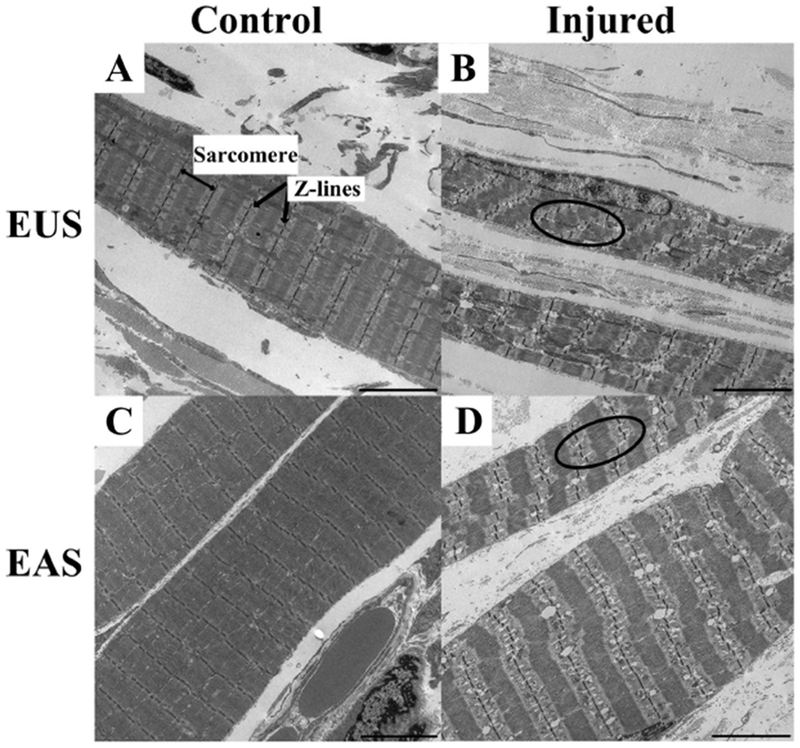
Aims: To define the operational resting sarcomere length (Ls ) of the female rat external urethral sphincter (EUS) and external anal sphincter (EAS) and to determine the mechanism of parturition-related injury of EUS and EAS using a simulated birth injury (SBI) vaginal distention model.
Methods: EUS and EAS of 3-month-old Sprague-Dawley control and injured rats were fixed in situ, harvested, and microdissected for Ls measurements and assessment of ultrastructure. EUS and EAS function was determined at baseline, and immediately and 4 weeks after SBI, using leak point pressure (LPP) and anorectal manometry (ARM), respectively. Operational L s was compared to species-specific optimal L s using one sample Student's t test. Data (mean ± SD) were compared between groups and time points using repeated measures one-way analysis of variance, followed by Tukey's post hoc pairwise comparisons, with significance set to 0.05.
Results: The operational resting Ls of both sphincters (EUS: 2.09 ± 0.07 µm, EAS: 2.02 ± 0.03 µm) was significantly shorter than optimal rat Ls of 2.4 µm. Strains imposed on EUS and EAS during SBI resulted in significant sarcomere elongation and disruption, compared with the controls (EUS: 3.09 ± 0.11 µm, EAS: 3.37 ± 0.09 µm). Paralleling structural changes, LPP and ARM measures were significantly lower immediately (LPP: 21.5 ± 1.0 cmH2 O, ARM: 5.1 ± 2.31 cmH2 O) and 4 weeks (LPP: 27.7 ± 1.3cmH2 O, ARM: 2.5 ± 1.0 cmH2 O) after SBI relative to the baseline (LPP: 43.4 ± 8.5 cmH2 O, ARM: 8.2 ± 2.0 cmH2 O); P < 0.05.
Conclusions: Analogous to humans, the short resting Ls of rat EUS and EAS favors their sphincteric function. The insult experienced by these muscles during parturition leads to sarcomere hyperelongation, myofibrillar disruption, and dysfunction of the sphincters long-term.
Keywords: birth injury; external anal sphincter; external urethral sphincter; fecal incontinence; rat; stress urinary incontinence.
© 2019 Wiley Periodicals, Inc.
Figures


Similar articles
-
Pelvic muscles' mechanical response to strains in the absence and presence of pregnancy-induced adaptations in a rat model.Am J Obstet Gynecol. 2018 May;218(5):512.e1-512.e9. doi: 10.1016/j.ajog.2018.02.001. Epub 2018 Feb 9. Am J Obstet Gynecol. 2018. PMID: 29432755 Free PMC article.
-
Long-term function and morphology of the anal sphincters and the pelvic floor after primary repair of obstetric anal sphincter injury.Colorectal Dis. 2014 Oct;16(10):O347-55. doi: 10.1111/codi.12579. Colorectal Dis. 2014. PMID: 24502361
-
Sustained improvement in the anal sphincter function following surgical plication of rabbit external anal sphincter muscle.Dis Colon Rectum. 2011 Nov;54(11):1373-80. doi: 10.1097/DCR.0b013e31822d0333. Dis Colon Rectum. 2011. PMID: 21979181
-
[Anorectal functional study. The state of the art].Minerva Chir. 1994 Dec;49(12):1187-93. Minerva Chir. 1994. PMID: 7746437 Review. Italian.
-
Neurogenic aspects of stress urinary incontinence.Curr Opin Obstet Gynecol. 2010 Oct;22(5):425-9. doi: 10.1097/GCO.0b013e32833e499d. Curr Opin Obstet Gynecol. 2010. PMID: 20706117 Free PMC article. Review.
Cited by
-
Sexual Dimorphism in the Architectural Design of Rat and Human Pelvic Floor Muscles.J Biomech Eng. 2024 Oct 1;146(10):101012. doi: 10.1115/1.4066090. J Biomech Eng. 2024. PMID: 39082779
References
Publication types
MeSH terms
Grants and funding
LinkOut - more resources
Full Text Sources
Research Materials
