. 2019 Apr 16;91(8):5106-5115.
doi: 10.1021/acs.analchem.8b05616. Epub 2019 Feb 22.

Multiplexed Relative Quantitation with Isobaric Tagging Mass Spectrometry Reveals Class I Major Histocompatibility Complex Ligand Dynamics in Response to Doxorubicin

Affiliations

Multiplexed Relative Quantitation with Isobaric Tagging Mass Spectrometry Reveals Class I Major Histocompatibility Complex Ligand Dynamics in Response to Doxorubicin

J Patrick Murphy et al. Anal Chem. .

Abstract

MHC-I peptides are intracellular-cleaved peptides, usually 8-11 amino acids in length, which are presented on the cell surface and facilitate CD8+ T cell responses. Despite the appreciation of CD8+ T-cell antitumor immune responses toward improvement in patient outcomes, the MHC-I peptide ligands that facilitate the response are poorly described. Along these same lines, although many therapies have been recognized for their ability to reinvigorate antitumor CD8+ T-cell responses, whether these therapies alter the MHC-I peptide repertoire has not been fully assessed due to the lack of quantitative strategies. We develop a multiplexing platform for screening therapy-induced MHC-I ligands by employing tandem mass tags (TMTs). We applied this approach to measuring responses to doxorubicin, which is known to promote antitumor CD8+ T-cell responses during its therapeutic administration in cancer patients. Using both in vitro and in vivo systems, we show successful relative quantitation of MHC-I ligands using TMT-based multiplexing and demonstrate that doxorubicin induces MHC-I peptide ligands that are largely derived from mitotic progression and cell-cycle proteins. This high-throughput MHC-I ligand discovery approach may enable further explorations to understand how small molecules and other therapies alter MHC-I ligand presentation that may be harnessed for CD8+ T-cell-based immunotherapies.


Conflict of interest statement

The authors declare no competing financial interest.

Figures

Figure 1.
Figure 1.
Platform for relative quantitative and multiplexed MHC-I peptidome analysis with TMT. Experiments begin with 6 to 11 samples (1 to 2 g of tissue or cell pellet per sample), followed by lysis, immunoprecipitation with an MHC/HLA-specific antibody, washing, and acid elution. Optionally, prior to lysis, a small portion of the sample can be saved for matched proteomic or proteogenomic analysis. Peptides are purified directly using MWCO or SPE and desalted using a Stage tip. Accurate relative quantitation between samples is achieved by SPS-MS3 data acquisition, and maximal HLA peptide identification is achieved with a targeted search strategy (enabled by SpectMHC) based on tissue HLA types. Optionally, exome or RNASeq data can be integrated into the targeted database. Finally, summed reporter ion intensities (S/N) represent individual HLA peptide relative quantitation across samples.
Figure 2.
Figure 2.
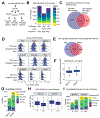
Effects of purification and TMT labeling on MHC-I peptidome composition. (A) Schematic to compare MWCO and SPE MHC-I peptide purification for TMT labeling in MDA MB 468 cells using TMT0. (B) Number of identified TMT0-labeled peptides by MWCO and SPE methods (both analyzed by SPS-MS3) compared with a standard MWCO and LC-MS2 method. (C) Overlap between TMT0-labeled and unlabeled HLA peptides (MWCO only). (D) Amino acid properties for MHC-I peptides found in the groups from panel C. (E) Overlap between MWCO- and SPE-purified TMT0-labeled HLA. (F) Comparison of logged signal-to-noise values of the TMT0 126 reporter ion between MWCO- and SPE-purified TMT0-labeled HLA peptides. (G) Quantifiable peptides (S/N > 100) in the TM0-labeled samples purified by either MWCO or SPE. (H) Mean logged S/N among peptides with or without the presence of lysine (K) in the sequence. (I) Quantifiable peptides in different S/N ranges among peptides with or without the presence of K in the sequence.
Figure 3.
Figure 3.
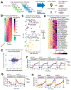
Dynamic, temporal induction of MHC-I peptides from chromatin and nucleosomal source proteins during time-course doxorubicin treatment of colon cancer cells. (A) Experimental schematic. HCT116 cells were treated over 24 h with 1 μM doxorubicin in duplicate. Cells were divided into MHC-I peptidome and proteome portions (90 and 10%, respectively) and subjected to MHC-I IP or tryptic digest, followed by multiplexed analyses with SPS-MS3. (B) Heatmap of global MHC-I peptide relative abundance over time. (C) Enriched GO terms (both Biological Process and Cellular Compartment) highlighting nuclear associations among doxorubicin-induced proteins. (D) Heatmap showing examples of highly induced MHC-I peptides from chromatin and chromosome segregation proteins. (E) Overall correlation between the mean relative peptide and matching mean source protein level changes after 24 h of doxorubicin treatment. (F) Examples of doxorubicin-induced MHC-I peptides from several nuclear-associated proteins (with no measurable change in the source proteins). (G) Examples of doxorubicin-repressed MHC-I peptides (associated with vesicle trafficking). (H) MHC-I peptides induced at the peptide and source levels, including two MHC-I peptides from MDM2 and one from KLHL21, both cell-cycle-related ubiquitin E3 ubiquitin ligases. Log Adj. pval = Benjamini–Hochberg-corrected F-test p value.
Figure 4.
Figure 4.
MHC-I peptide multiplexing in a mouse tumor model of doxorubicin treatment. (A) Experimental setup. Tumors (EL4 lymphoma) and spleens (n = 2) from mice treated or untreated with 2.5 mg/kg doxorubicin or vehicle (PBS) were harvested and subjected to multiplexed MHC-I peptidome or proteome analysis. Cultured EL4 cells (doxorubicin- or PBS-treated, n = 1) were also included as a comparison. (B) Volcano plot of doxorubicin effects on the MHC-I peptides in the EL4 tumors and spleens from the same mice. (C) Heatmaps of doxorubicin-induced MHC-I peptides from both the tumor (left) and the spleen (right) as they appear across all 10 samples. (D) Strategy for matching MHC-I peptides to potential source proteins. (E) Number of MHC-I peptides (with H-2 Kb or H-2 Db specificity) matching the multiplexed proteome data set. (F) Correlation of doxorubicin-induced changes in both the tumor (left) and spleen (right) at the MHC-I peptide and source protein levels.
Figure 5.
Figure 5.
Tissue specificity in relative quantitative MHC-I peptidome data from the EL4 mouse tumor model with doxorubicin. (A) Principal component analysis (PCA) of multiplexed MHC-I peptide data in the EL4 tumor model experiment. (B) Heatmap of MHC-I peptides (3034 unique peptides) specific to spleen, tumor, or in vitro tumors, independent of doxorubicin treatment. (C) Sequence logos demonstrating upstream and downstream proteasomal cleavage and trimming specificity of tumor- and spleen-specific MHC-I peptides (compared with nonspecific peptides). (D) Differential frequency of amino acids in the upstream and downstream sequences of spleen- and tumor-specific MHC-I peptides.

References

    1. Matsushita H; Vesely MD; Koboldt DC; Rickert CG; Uppaluri R; Magrini VJ; Arthur CD; White JM; Chen Y-S; Shea LK; et al. Nature 2012, 482 (7385), 400–404. - PMC - PubMed
    1. Castle JC; Kreiter S; Diekmann J; Lower M; van de Roemer N; de Graaf J; Selmi A; Diken M; Boegel S; Paret C; et al. Cancer Res. 2012, 72 (5), 1081–1091. - PubMed
    1. Yadav M; Jhunjhunwala S; Phung QT; Lupardus P; Tanguay J; Bumbaca S; Franci C; Cheung TK; Fritsche J; Weinschenk T; et al. Nature 2014, 515 (7528), 572–576. - PubMed
    1. Lemmel C; Weik S; Eberle U; Dengjel J; Kratt T; Becker H-D; Rammensee H-G; Stevanović S Nat. Biotechnol 2004, 22 (4), 450–454. - PubMed
    1. Cox J; Hein MY; Luber CA; Paron I; Nagaraj N; Mann M Mol. Cell. Proteomics 2014, 13 (9), 2513–2526. - PMC - PubMed

Publication types

MeSH terms