

Circulating Microparticles Are Elevated in Treated HIV -1 Infection and Are Deleterious to Endothelial Cell Function
- PMID: 30779672
- PMCID: PMC6405669
- DOI: 10.1161/JAHA.118.011134
Circulating Microparticles Are Elevated in Treated HIV -1 Infection and Are Deleterious to Endothelial Cell Function
Abstract
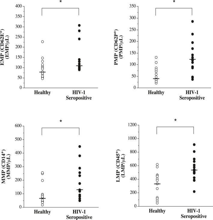
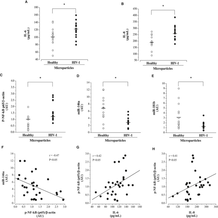
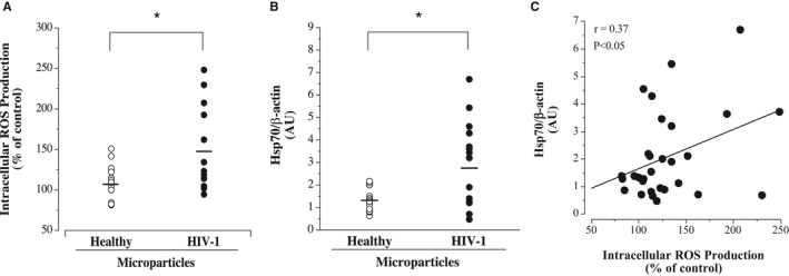
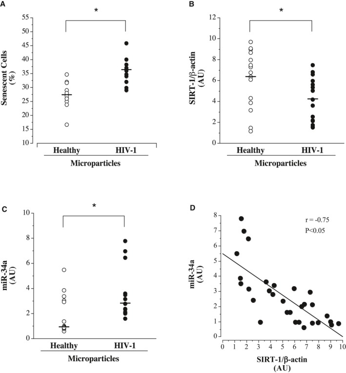
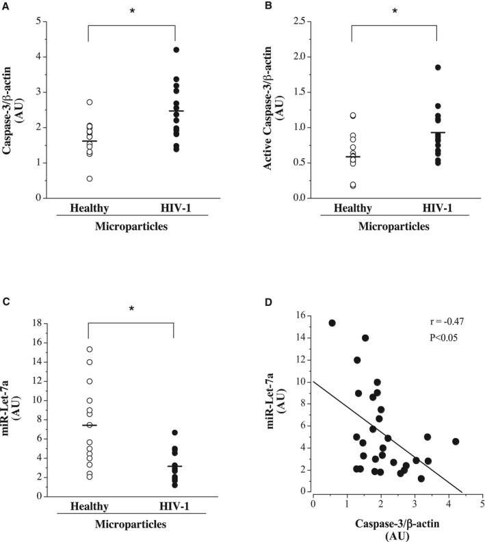
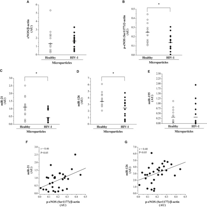
Background Circulating microparticles have emerged as biomarkers and effectors of vascular disease. Elevated rates of cardiovascular disease are seen in HIV -1-seropositive individuals. The aims of this study were to determine: (1) if circulating microparticles are elevated in antiretroviral therapy-treated HIV -1-seropositive adults; and (2) the effects of microparticles isolated from antiretroviral therapy -treated HIV -1-seropositive adults on endothelial cell function, in vitro. Methods and Results Circulating levels of endothelial-, platelet-, monocyte-, and leukocyte-derived microparticles were determined by flow cytometry in plasma from 15 healthy and 15 antiretroviral therapy-treated, virologically suppressed HIV -1-seropositive men. Human umbilical vein endothelial cells were treated with microparticles from individual subjects for 24 hours; thereafter, endothelial cell inflammation, oxidative stress, senescence, and apoptosis were assessed. Circulating concentrations of endothelial-, platelet-, monocyte-, and leukocyte-derived microparticles were significantly higher (≈35%-225%) in the HIV -1-seropositive compared with healthy men. Microparticles from HIV -1-seropositive men induced significantly greater endothelial cell release of interleukin-6 and interleukin-8 (≈20% and ≈35%, respectively) and nuclear factor-κB expression while suppressing anti-inflammatory microRNAs (miR-146a and miR-181b). Intracellular reactive oxygen species production and expression of reactive oxygen species -related heat shock protein 70 were both higher in cells treated with microparticles from the HIV -1-seropositive men. In addition, the percentage of senescent cells was significantly higher and sirtuin 1 expression lower in cells treated with HIV -1-related microparticles. Finally, caspase-3 was significantly elevated by microparticles from HIV -1-seropositive men. Conclusions Circulating concentrations of endothelial-, platelet-, monocyte-, and leukocyte-derived microparticles were higher in antiretroviral therapy-treated HIV -1-seropositive men and adversely affect endothelial cells promoting cellular inflammation, oxidative stress, senescence, and apoptosis. Circulating microparticles may contribute to the vascular risk associated with HIV -1 infection.
Keywords: HIV‐1; endothelial dysfunction; inflammation; microRNA; microparticles.
Figures
Similar articles
-
Imbalance between endothelial progenitors cells and microparticles in HIV-infected patients naive for antiretroviral therapy.AIDS. 2011 Aug 24;25(13):1595-601. doi: 10.1097/QAD.0b013e32834980f4. AIDS. 2011. PMID: 21673561
-
Effects of HIV-1 gp120 and TAT-derived microvesicles on endothelial cell function.J Appl Physiol (1985). 2019 May 1;126(5):1242-1249. doi: 10.1152/japplphysiol.01048.2018. Epub 2019 Feb 21. J Appl Physiol (1985). 2019. PMID: 30789287 Free PMC article.
-
Influence of sex on the number of circulating endothelial microparticles and microRNA expression in middle-aged adults.Exp Physiol. 2017 Aug 1;102(8):894-900. doi: 10.1113/EP086359. Epub 2017 Jun 30. Exp Physiol. 2017. PMID: 28568648
-
Endothelial dysfunction in HIV infection--the role of circulating endothelial cells, microparticles, endothelial progenitor cells and macrophages.AIDS Rev. 2012 Oct-Dec;14(4):223-30. AIDS Rev. 2012. PMID: 23258297 Review.
-
Microparticles are vectors of paradoxical information in vascular cells including the endothelium: role in health and diseases.Pharmacol Rep. 2008 Jan-Feb;60(1):75-84. Pharmacol Rep. 2008. PMID: 18276988 Review.
Cited by
-
Circulating extracellular microvesicles associated with electronic cigarette use increase endothelial cell inflammation and reduce nitric oxide production.Exp Physiol. 2024 Sep;109(9):1593-1603. doi: 10.1113/EP091715. Epub 2024 Aug 2. Exp Physiol. 2024. PMID: 39092897 Free PMC article.
-
Neuropilin-1 in the pathogenesis of preeclampsia, HIV-1, and SARS-CoV-2 infection: A review.Virus Res. 2022 Oct 2;319:198880. doi: 10.1016/j.virusres.2022.198880. Epub 2022 Jul 26. Virus Res. 2022. PMID: 35905790 Free PMC article. Review.
-
Plasma Extracellular Vesicles Enhance HIV-1 Infection of Activated CD4+ T Cells and Promote the Activation of Latently Infected J-Lat10.6 Cells via miR-139-5p Transfer.Front Immunol. 2021 Jun 24;12:697604. doi: 10.3389/fimmu.2021.697604. eCollection 2021. Front Immunol. 2021. PMID: 34249000 Free PMC article.
-
Novel Mechanism of Microvesicle Regulation by the Antiviral Protein Tetherin During HIV Infection.J Am Heart Assoc. 2020 Sep;9(17):e015998. doi: 10.1161/JAHA.120.015998. Epub 2020 Aug 21. J Am Heart Assoc. 2020. PMID: 32819189 Free PMC article.
-
Circulating Cell Biomarkers in Pulmonary Arterial Hypertension: Relationship with Clinical Heterogeneity and Therapeutic Response.Cells. 2021 Jul 4;10(7):1688. doi: 10.3390/cells10071688. Cells. 2021. PMID: 34359858 Free PMC article.
References
-
- Freiberg MS, Chang C‐CH, Kuller LH, Skanderson M, Lowy E, Kraemer KL, Butt AA, Bidwell Goetz M, Leaf D, Oursler KA, Rimland D, Rodriguez Barradas M, Brown S, Gibert C, McGinnis K, Crothers K, Sico J, Crane H, Warner A, Gottlieb S, Gottdiener J, Tracy RP, Budoff M, Watson C, Armah KA, Doebler D, Bryant K, Justice AC. HIV infection and the risk of acute myocardial infarction. JAMA Intern Med. 2013;173:614–622. - PMC - PubMed
-
- Islam FM, Wu J, Jansson J, Wilson DP. Relative risk of cardiovascular disease among people living with HIV: a systematic review and meta‐analysis. HIV Med. 2012;13:453–468. - PubMed
-
- Gibellini D, Borderi M, Clò A, Morini S, Miserocchi A, Bon I, Ponti C, Re MC. HIV‐related mechanisms in atherosclerosis and cardiovascular diseases. J Cardiovasc Med (Hagerstown). 2013;14:780–790. - PubMed
-
- Libby P, Theroux P. Pathophysiology of coronary artery disease. Circulation. 2005;111:3481–3488. - PubMed
Publication types
MeSH terms
Substances
Grants and funding
LinkOut - more resources
Full Text Sources
Medical
Research Materials
