

Osseointegration of Alkali-Modified NANOZR Implants: An In Vivo Study
- PMID: 30781372
- PMCID: PMC6413168
- DOI: 10.3390/ijms20040842
Osseointegration of Alkali-Modified NANOZR Implants: An In Vivo Study
Abstract
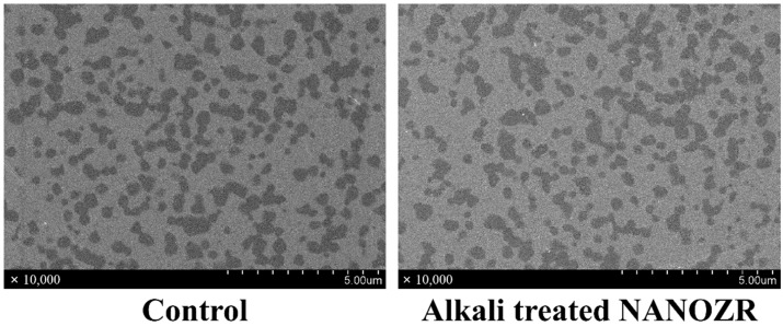
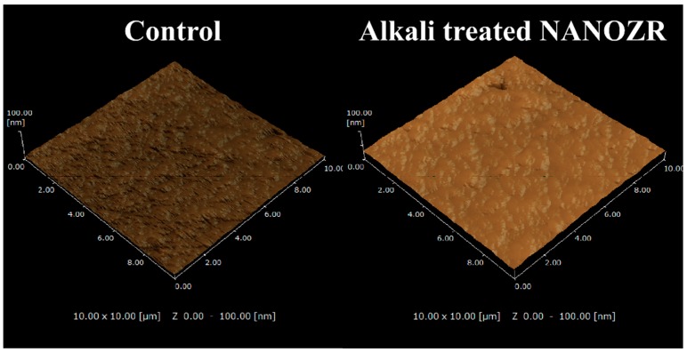
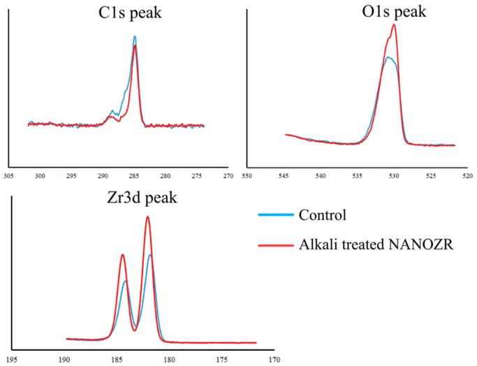
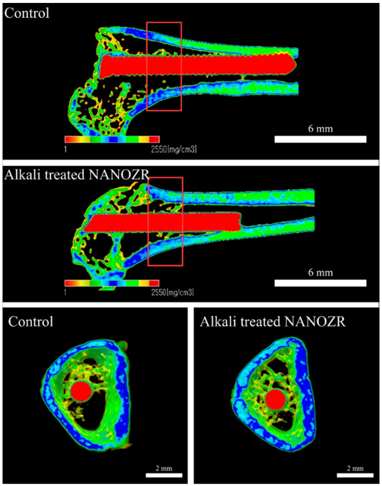
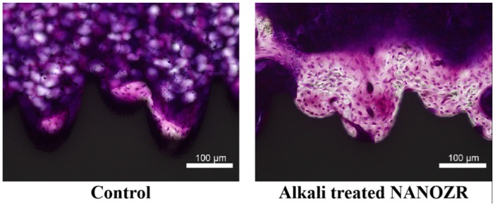
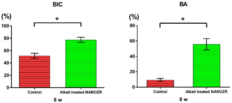
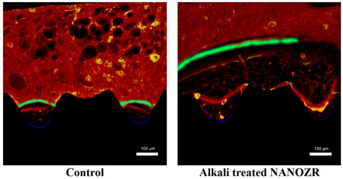
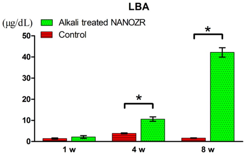
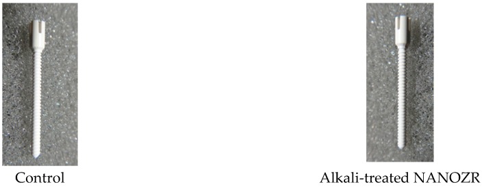
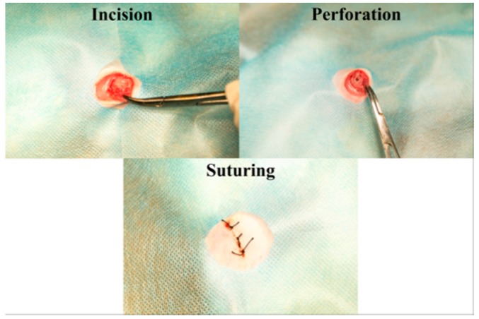
Ingredients and surface modification methods are being continually developed to improve osseointegration of dental implants and reduce healing times. In this study, we demonstrate in vitro that, by applying concentrated alkali treatment to NANOZR with strong bending strength and fracture toughness, a significant improvement in the bone differentiation of rat bone marrow cells can be achieved. We investigated the influence of materials modified with this treatment in vivo, on implanted surrounding tissues using polychrome sequential fluorescent labeling and micro-computer tomography scanning. NANOZR implant screws in the alkali-treated group and the untreated group were evaluated after implantation in the femur of Sprague⁻Dawley male rats, indicating that the amount of new bone in the alkali-modified NANOZR was higher than that of unmodified NANOZR. Alkali-modified NANOZR implants proved to be useful for the creation of new implant materials.
Keywords: NANOZR; alkali treatment; in vivo study.
Conflict of interest statement
The authors declare no conflict of interest.
Figures

Similar articles
-
Effects of Plasma Treatment on the Bioactivity of Alkali-Treated Ceria-Stabilised Zirconia/Alumina Nanocomposite (NANOZR).Int J Mol Sci. 2020 Oct 10;21(20):7476. doi: 10.3390/ijms21207476. Int J Mol Sci. 2020. PMID: 33050494 Free PMC article.
-
Bioactivity of NANOZR Induced by Alkali Treatment.Int J Mol Sci. 2017 Apr 6;18(4):780. doi: 10.3390/ijms18040780. Int J Mol Sci. 2017. PMID: 28383491 Free PMC article.
-
Biomechanical and histological evaluation of the osseointegration capacity of two types of zirconia implant.Int J Nanomedicine. 2016 Dec 7;11:6507-6516. doi: 10.2147/IJN.S119519. eCollection 2016. Int J Nanomedicine. 2016. PMID: 27994456 Free PMC article.
-
Osseointegration of titanium, titanium alloy and zirconia dental implants: current knowledge and open questions.Periodontol 2000. 2017 Feb;73(1):22-40. doi: 10.1111/prd.12179. Periodontol 2000. 2017. PMID: 28000277 Review.
-
Peri-implant osteogenesis in health and osteoporosis.Micron. 2005;36(7-8):630-44. doi: 10.1016/j.micron.2005.07.008. Epub 2005 Sep 6. Micron. 2005. PMID: 16182543 Review.
Cited by
-
Effects of Plasma Treatment on the Bioactivity of Alkali-Treated Ceria-Stabilised Zirconia/Alumina Nanocomposite (NANOZR).Int J Mol Sci. 2020 Oct 10;21(20):7476. doi: 10.3390/ijms21207476. Int J Mol Sci. 2020. PMID: 33050494 Free PMC article.
-
Effect of Plasma Treatment on Titanium Surface on the Tissue Surrounding Implant Material.Int J Mol Sci. 2021 Jun 28;22(13):6931. doi: 10.3390/ijms22136931. Int J Mol Sci. 2021. PMID: 34203231 Free PMC article.
-
Nanostructured Zirconia-Based Ceramics and Composites in Dentistry: A State-of-the-Art Review.Nanomaterials (Basel). 2019 Sep 29;9(10):1393. doi: 10.3390/nano9101393. Nanomaterials (Basel). 2019. PMID: 31569589 Free PMC article. Review.
-
An engineering perspective of ceramics applied in dental reconstructions.J Appl Oral Sci. 2023 Feb 20;31:e20220421. doi: 10.1590/1678-7757-2022-0421. eCollection 2023. J Appl Oral Sci. 2023. PMID: 36820784 Free PMC article. Review.
-
Effect of Argon-Based Atmospheric Pressure Plasma Treatment on Hard Tissue Formation on Titanium Surface.Int J Mol Sci. 2021 Jul 16;22(14):7617. doi: 10.3390/ijms22147617. Int J Mol Sci. 2021. PMID: 34299241 Free PMC article.
References
-
- Adell R., Eriksson B., Lekholm U. A long-term follow up study of osseointegrated implants in the treatment of totally edentulous jaws. Int. J. Oral Maxillofac. Implants. 1990;5:347–359. - PubMed
-
- Henry P.J., Laney W.R., Jemt T. Osseointegrated implants for single-tooth replacement: A prospective 5-year multicenter study. Int. J. Oral Maxillofac. Implants. 1996;11:450–455. - PubMed
MeSH terms
Substances
LinkOut - more resources
Full Text Sources
