Review
. 2019 Feb 15;11(2):226.
doi: 10.3390/cancers11020226.

Detecting Chromosome Instability in Cancer: Approaches to Resolve Cell-to-Cell Heterogeneity

Affiliations
Review

Detecting Chromosome Instability in Cancer: Approaches to Resolve Cell-to-Cell Heterogeneity

Chloe C Lepage et al. Cancers (Basel). .

Abstract

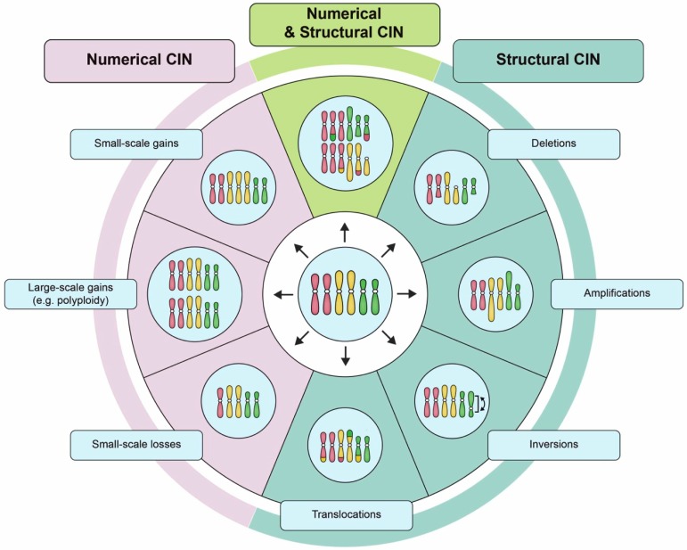
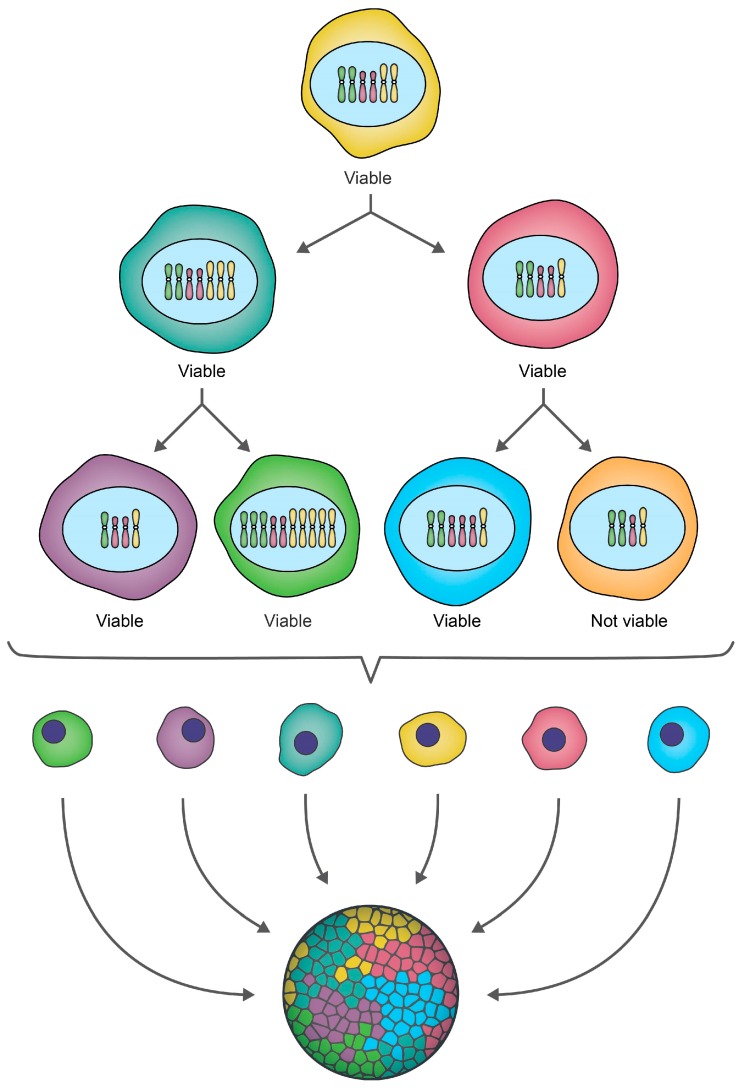
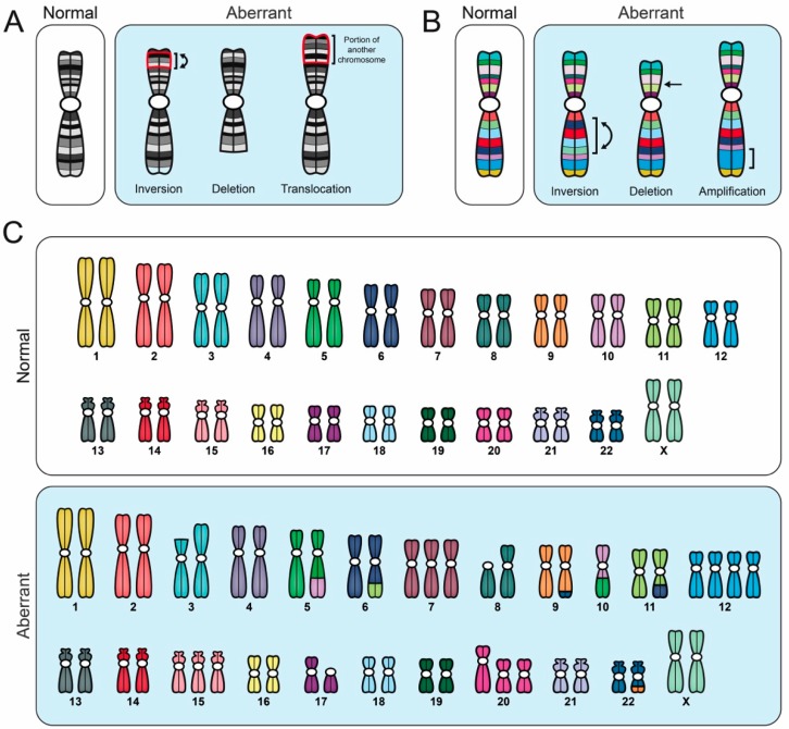
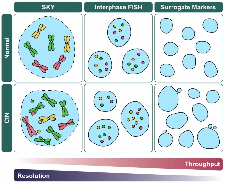
Chromosome instability (CIN) is defined as an increased rate of chromosome gains and losses that manifests as cell-to-cell karyotypic heterogeneity and drives cancer initiation and evolution. Current research efforts are aimed at identifying the etiological origins of CIN, establishing its roles in cancer pathogenesis, understanding its implications for patient prognosis, and developing novel therapeutics that are capable of exploiting CIN. Thus, the ability to accurately identify and evaluate CIN is critical within both research and clinical settings. Here, we provide an overview of quantitative single cell approaches that evaluate and resolve cell-to-cell heterogeneity and CIN, and discuss considerations when selecting the most appropriate approach to suit both research and clinical contexts.

Keywords: cancer; chromosome instability; intratumoral heterogeneity; quantitative imaging microscopy; single cell approaches.


Conflict of interest statement

The authors declare no conflict of interest.

Figures

Figure 1
Figure 1
Examples of numerical chromosome instability (CIN) and structural CIN. A schematic depicting examples of the types of karyotypic changes associated with either numerical CIN (N-CIN) or structural CIN (S-CIN). Note that to accurately define CIN within a given population requires multiple distinct karyotypes to be identified as a single aberrant karyotype only defines a state, and not a rate. For illustrative purposes, the starting diploid cell (center) only contains three pairs of chromosomes (i.e., a partial karyotype). N-CIN involves whole chromosome gains or losses, including both small-scale changes that result in aneuploidy, as well as large-scale polyploidization events. S-CIN includes partial chromosome deletions, amplifications, inversions, or translocations (ranging in size from single genes to entire chromosome arms). These different classes of N-CIN or S-CIN are often combined to produce complex karyotypes that evolve over time. However, techniques for evaluating CIN typically only detect a subset of these karyotypic changes.
Figure 2
Figure 2
CIN drives ongoing karyotypic heterogeneity within cellular populations. A schematic depicting a hypothetical example of CIN within an initial cell that for illustrative purposes contains only three pairs of chromosomes (i.e., a partial karyotype). As this cell undergoes two rounds of cellular division, chromosomes are gained or lost, producing a heterogeneous population of genetically distinct daughter cells, which is referred to as intratumoral heterogeneity in a cancer context. Some karyotypic changes may not be compatible with cell viability, as indicated by the orange cell which is lost from the population. Note that while this example focuses on small-scale gains/losses of whole chromosomes (N-CIN), chromosome complements may also evolve via increases in ploidy (N-CIN) or structural chromosome changes (S-CIN) (see Figure 1), and often include a combination of both N- and S-CIN.
Figure 3
Figure 3
Comparison of different cytogenetic techniques used to evaluate CIN. Schematic illustrating various aberrant karyotypic events detected using specific cytogenetic techniques, all of which are performed on mitotic chromosome spreads. Importantly, to accurately identify CIN (i.e., cell-to-cell heterogeneity), numerous distinct aberrant events must be identified within a given population; a single aberrant karyotype that is stable within the population does not constitute CIN. (A) G-banding and inverted DAPI counterstaining enable full karyotypic assessment, and they identify individual chromosomes based on their unique banding pattern. Aberrant banding patterns are suggestive of structural abnormalities (i.e., S-CIN). (B) Multiplex-banding (M-banding) fluorescence in situ hybridization (FISH) employs multicolored probes generated for a specific chromosome or region, and they can be used to identify aberrant intrachromosomal events like inversions, deletions or amplifications (i.e., S-CIN). (C) Spectral karyotyping (SKY) employs multicolored probes to ‘paint’ each chromosome a unique color, enabling a full karyotypic assessment. SKY can detect chromosome copy number changes (gains or losses), interchromosomal translocations and some intrachromosomal events (large deletions and amplifications), but does not readily detect intrachromosomal inversions.
Figure 4
Figure 4
CIN-based analytics and the balance between data throughput and resolution. Schematic illustrating various quantitative imaging approaches used to assess CIN and their strengths in data throughput versus resolution (i.e., N- versus S-CIN). For illustrative purposes, representative examples of normal (top) and CIN-positive (bottom; N- and S-CIN) cellular contexts are shown. FISH (Section 4.2) and related approaches (e.g., fluorescent operator/reporter systems; Section 3.2) rely on enumeration of fluorescent foci rather than whole chromosomes which facilitates manual or automated analyses, but they do not detect N- or S-CIN involving other loci. Spectral karyotyping (SKY; Section 4.3) enables a genome-wide assessment of N- and S-CIN, but is not typically used to assess large sample sizes. Surrogate markers of CIN including changes in nuclear area and micronucleus formation (Section 5.1), are amenable to high-throughput approaches, but they do not specifically assess N- or S-CIN, and they may be subject to false positive/negative results.

References

    1. Hanahan D., Weinberg R.A. Hallmarks of cancer: The next generation. Cell. 2011;144:646–674. doi: 10.1016/j.cell.2011.02.013. - DOI - PubMed
    1. Geigl J.B., Obenauf A.C., Schwarzbraun T., Speicher M.R. Defining “chromosomal instability”. Trends Genet. 2008;24:64–69. doi: 10.1016/j.tig.2007.11.006. - DOI - PubMed
    1. Sieber O.M., Heinimann K., Tomlinson I. Genomic instability—The engine of tumorigenesis? Nat. Rev. Cancer. 2003;3:469–476. doi: 10.1038/nrc1170. - DOI - PubMed
    1. Beckman R.A., Loeb L.A. Genetic instability in cancer: Theory and experiment. Semin. Cancer Biol. 2005;15:423–435. doi: 10.1016/j.semcancer.2005.06.007. - DOI - PubMed
    1. Rajagopalan H., Nowak M.A., Vogelstein B. The significance of unstable chromosomes in colorectal cancer. Nat. Rev. Cancer. 2003;3:5–10. doi: 10.1038/nrc1165. - DOI - PubMed

LinkOut - more resources