. 2019 Feb 15;20(4):854.
doi: 10.3390/ijms20040854.

o-Vanillin Derived Schiff Bases and Their Organotin(IV) Compounds: Synthesis, Structural Characterisation, In-Silico Studies and Cytotoxicity

Affiliations

o-Vanillin Derived Schiff Bases and Their Organotin(IV) Compounds: Synthesis, Structural Characterisation, In-Silico Studies and Cytotoxicity

Enis Nadia Md Yusof et al. Int J Mol Sci. .

Abstract

Six new organotin(IV) compounds of Schiff bases derived from S-R-dithiocarbazate [R = benzyl (B), 2- or 4-methylbenzyl (2M and 4M, respectively)] condensed with 2-hydroxy-3-methoxybenzaldehyde (oVa) were synthesised and characterised by elemental analysis, various spectroscopic techniques including infrared, UV-vis, multinuclear (¹H, 13C, 119Sn) NMR and mass spectrometry, and single crystal X-ray diffraction. The organotin(IV) compounds were synthesised from the reaction of Ph₂SnCl₂ or Me₂SnCl₂ with the Schiff bases (S2MoVaH/S4MoVaH/SBoVaH) to form a total of six new organotin(IV) compounds that had a general formula of [R₂Sn(L)] (where L = Schiff base; R = Ph or Me). The molecular geometries of Me₂Sn(S2MoVa), Me₂Sn(S4MoVa) and Me₂Sn(SBoVa) were established by X-ray crystallography and verified using density functional theory calculations. Interestingly, each experimental structure contained two independent but chemically similar molecules in the crystallographic asymmetric unit. The coordination geometry for each molecule was defined by thiolate-sulphur, phenoxide-oxygen and imine-nitrogen atoms derived from a dinegative, tridentate dithiocarbazate ligand with the remaining positions occupied by the methyl-carbon atoms of the organo groups. In each case, the resulting five-coordinate C₂NOS geometry was almost exactly intermediate between ideal trigonal-bipyramidal and square-pyramidal geometries. The cytotoxic activities of the Schiff bases and organotin(IV) compounds were investigated against EJ-28 and RT-112 (bladder), HT29 (colon), U87 and SJ-G2 (glioblastoma), MCF-7 (breast) A2780 (ovarian), H460 (lung), A431 (skin), DU145 (prostate), BE2-C (neuroblastoma) and MIA (pancreatic) cancer cell lines and one normal breast cell line (MCF-10A). Diphenyltin(IV) compounds exhibited greater potency than either the Schiff bases or the respective dimethyltin(IV) compounds. Mechanistic studies on the action of these compounds against bladder cancer cells revealed that they induced the production of reactive oxygen species (ROS). The bladder cancer cells were apoptotic after 24 h post-treatment with the diphenyltin(IV) compounds. The interactions of the organotin(IV) compounds with calf thymus DNA (CT-DNA) were experimentally explored using UV-vis absorption spectroscopy. This study revealed that the organotin(IV) compounds have strong DNA binding affinity, verified via molecular docking simulations, which suggests that these organotin(IV) compounds interact with DNA via groove-binding interactions.

Keywords: cytotoxic activity; dithiocarbazate; five-coordinate compounds; mechanistic studies; molecular docking; organotin(IV); single-crystal X-ray diffraction analysis; tridentate ONS Schiff bases.


Conflict of interest statement

The authors declare no conflict of interest.

Figures

Scheme 1
Scheme 1
Synthetic pathway for the formation of SBoVaH, S2MoVaH, and S4MoVaH.
Scheme 2
Scheme 2
Synthesis of organotin(IV) compounds.
Figure 1
Figure 1
The thione (a) and thiol (b) tautomeric forms of the Schiff bases (R = o–CH3, S2MoVaH; p-CH3, S4MoVaH; R = H, SBoVaH).
Figure 2
Figure 2
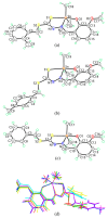
Molecular structures of the first independent molecules of (a) Me2Sn(S2MoVa), (b) Me2Sn(S4MoVa) and (c) Me2Sn(SBoVa) showing atom labelling schemes. (d) Overlay diagram of the two independent molecules of each of Me2Sn(S2MoVa), a (red image) and b (green), Me2Sn(S4MoVa), a (blue) and b (pink), and Me2Sn(SBoVa) a (yellow) and b (aqua) with the SnMe2 planes superimposed.
Figure 3
Figure 3
Crystallographic diagrams for Me2Sn(S2MoVa): (a) Molecular structure of the second independent molecule, molecule b, (b) overlay diagram of molecules a (red image) and inverted-b (green) drawn so the SnC2 atoms of the Me2Sn moiety are overlapped and (c) a view of the supramolecular layer in the ab-plane (left image) and a view of the unit cell contents in projection down the a-axis with one layer highlighted in space-filling mode (right image). The C–HO and C–Hπ interactions are shown as orange and purple dashed lines, respectively.
Figure 4
Figure 4
Crystallographic diagrams for Me2Sn(S4MoVa): (a) Molecular structure of the second independent molecule, molecule b, (b) overlay diagram of molecules a (blue image) and inverted-b (pink) drawn so the SnC2 atoms of the Me2Sn moiety are overlapped and (c) a view of the supramolecular dimer sustained by C–Hπ (chelate) interactions (left image; non-participating hydrogen atoms have been omitted) and a view of the unit cell contents in projection down the a-axis. The C–HO and C–Hπ interactions are shown as orange and purple dashed lines, respectively.
Figure 5
Figure 5
Crystallographic diagrams for Sn(SBoVa)Me2: (a) Molecular structure of the second independent molecule, molecule b, (b) overlay diagram of molecules a (yellow image) and b (aqua) drawn so the five-membered rings are overlapped and (c) supramolecular dimer sustained by edge-to-edge chelate ringbenzene interactions (upper image) between Sn1-containing molecules, supramolecular layer sustained by edge-to-edge π (chelate ring)π (ethoxybenzene) and phenyl-C–Hπ (methoxybenzene) interactions occurring between Sn2-containing molecules and a view of the unit cell contents in projection down the b-axis. The edge-to-edge π (chelate ring)π (ethoxybenzene) and C–Hπ interactions are shown as blue and purple dashed lines, respectively.
Figure 6
Figure 6
Apoptosis detection through fluorescence microscopy. Cells were treated for 24 h with Ph2Sn(SBoVa) (0.58 µM), Ph2Sn(S2MoVa) (0.31 µM) and Ph2Sn(S4MoVa) (1.66 µM) and the negative control (DMSO) in complete media. After staining with Annexin V and PI, necrotic and apoptotic cells were detected by fluorescence microscopy (20×).
Figure 7
Figure 7
Percent reactive oxygen species (ROS) production in RT-112 cells treated with (a) Ph2Sn(S2MoVa) (0.31 μM) (b) Ph2Sn(S4MoVa) (1.66 μM) for 24 h and stained with 1 mM DCFH-DA for 60 minutes at 37 °C. DMSO and H2O2 acted as negative and positive controls, respectively.
Figure 8
Figure 8
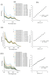
(a) Electronic absorption spectra of (i) Ph2Sn(S2MoVa), (ii) Ph2Sn(S4MoVa) and (iii) Ph2Sn(SBoVa); (b) Plot of [DNA]/εa − εf vs. [DNA] for absorption titration of DNA with (i) Ph2Sn(S2MoVa), (ii) Ph2Sn(S4MoVa) and (iii) Ph2Sn(SBoVa). The arrow indicates the change in absorbance in tandem with increasing DNA concentration.
Figure 9
Figure 9
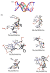
(a) Schematic representation of organotin(IV) compounds that fit well in the grooves of the DNA strand obtained by docking simulations. The two double-stranded DNA comprise of the phosphate deoxyribose backbone with guanine (DG, red), cytosine (DC, blue), adenine (DA, pink) and thymine (DT, orange). (b) Molecular interactions of organotin(IV) compounds within the grooves of double stranded DNA residues.

References

    1. Boseley S. [(accessed on 20 February 2018)]; Available online: https://www.theguardian.com/society/2014/feb/03/worldwide-cancer-cases-s....
    1. Global Cancer Observatory. [(accessed on 20 February 2018)]; Available online: http://gco.iarc.fr/
    1. Lebwohl D., Canetta R. Clinical development of platinum complexes in cancer therapy: An historical perspective and an update. Eur. J. Cancer. 1998;34:1522–1534. doi: 10.1016/S0959-8049(98)00224-X. - DOI - PubMed
    1. Galanski M. Recent Developments in the Field of Anticancer Platinum Complexes. Recent Pat. Anticancer Drug Discov. 2006;1:285–295. doi: 10.2174/157489206777442287. - DOI - PubMed
    1. Sirajuddin M., Ali S., Mckee V., Zaib S., Iqbal J. Organotin(IV) carboxylate derivatives as a new addition to anticancer and antileishmanial agents: Design, physicochemical characterization and interaction with Salmon sperm DNA. RSC Adv. 2014;4:57505–57521. doi: 10.1039/C4RA10487K. - DOI

MeSH terms

LinkOut - more resources