. 2019 Feb 16;8(2):251.
doi: 10.3390/jcm8020251.

Biobanking: Objectives, Requirements, and Future Challenges-Experiences from the Munich Vascular Biobank

Affiliations

Biobanking: Objectives, Requirements, and Future Challenges-Experiences from the Munich Vascular Biobank

Jaroslav Pelisek et al. J Clin Med. .

Abstract

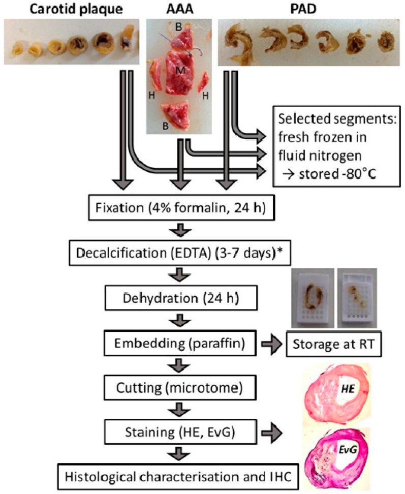
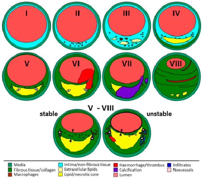
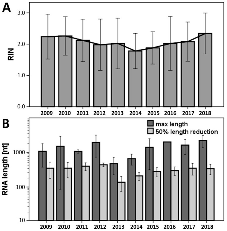
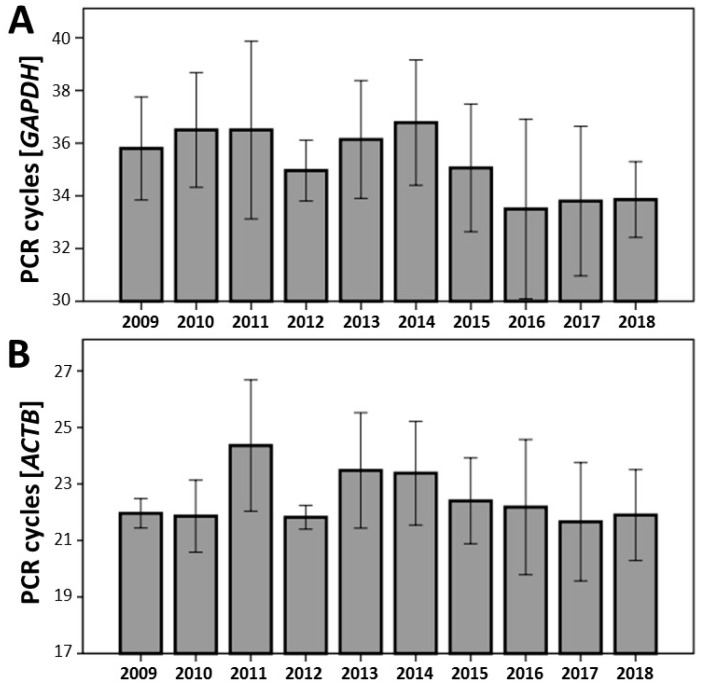
Collecting biological tissue samples in a biobank grants a unique opportunity to validate diagnostic and therapeutic strategies for translational and clinical research. In the present work, we provide our long-standing experience in establishing and maintaining a biobank of vascular tissue samples, including the evaluation of tissue quality, especially in formalin-fixed paraffin-embedded specimens (FFPE). Our Munich Vascular Biobank includes, thus far, vascular biomaterial from patients with high-grade carotid artery stenosis (n = 1567), peripheral arterial disease (n = 703), and abdominal aortic aneurysm (n = 481) from our Department of Vascular and Endovascular Surgery (January 2004⁻December 2018). Vascular tissue samples are continuously processed and characterized to assess tissue morphology, histological quality, cellular composition, inflammation, calcification, neovascularization, and the content of elastin and collagen fibers. Atherosclerotic plaques are further classified in accordance with the American Heart Association (AHA), and plaque stability is determined. In order to assess the quality of RNA from FFPE tissue samples over time (2009⁻2018), RNA integrity number (RIN) and the extent of RNA fragmentation were evaluated. Expression analysis was performed with two housekeeping genes-glyceraldehyde 3-phosphate dehydrogenase (GAPDH) and beta-actin (ACTB)-using TaqMan-based quantitative reverse-transcription polymerase chain reaction (qRT)-PCR. FFPE biospecimens demonstrated unaltered RNA stability over time for up to 10 years. Furthermore, we provide a protocol for processing tissue samples in our Munich Vascular Biobank. In this work, we demonstrate that biobanking is an important tool not only for scientific research but also for clinical usage and personalized medicine.

Keywords: Munich Vascular Biobank; RIN; RNA fragmentation; atherosclerosis; human vascular tissue.


Conflict of interest statement

The authors declare no conflict of interest.

Figures

Figure 1
Figure 1
Evaluation of the extent of RNA fragmentation from FFPE tissue samples measuring the area under the curve from Agilent Bioanalyzer. Two values were defined: maximal RNA length and the 50% RNA length calculated as a 50% reduction of the area under the curve. RIN: RNA integrity number; FFPE: formalin-fixed paraffin-embedded specimens. nt: number of nucleotides; FU: fluorescence unit.
Figure 2
Figure 2
Examples of tissue samples collected in our Munich Vascular Biobank. CAR: carotid atherosclerotic plaques obtained from patients with high-grade carotid artery stenosis (>50%) [16] by endarterectomy (EA); PAD: atherosclerotic plaques from patients with peripheral artery disease obtained by EA [17]; AAA: aortic wall from patients with abdominal aortic aneurysm who underwent open surgical repair [18]; B: fresh frozen segments for molecular biology; H: histology; M: mechanics (tensile tests); CT: computer tomography, MRI: magnetic resonance imaging.
Figure 3
Figure 3
Schematic chart of the processing of vascular tissue after surgical excision. Carotid plaque: atherosclerotic lesions from patients with high-graded carotid artery stenosis; AAA: aortic wall from patients with abdominal aortic aneurysm; PAD: atherosclerotic tissue from patients with peripheral artery disease; RT: room temperature; IHC: immunohistochemistry. HE: haematoxilin-eosin staining; EvG: elastica van gieson staining; * the time depends on the extent of calcification.
Figure 4
Figure 4
Classification of atherosclerotic lesions according to the American Heart Association (AHA) [24,25,26,27]. Type I: initial lesion with isolated macrophages and macrophage-derived foam cells; type II: fatty streaks, increased number of foam cells, intracellular lipid accumulation; type III: further accumulation of inflammatory cells and intracellular lipids, isolated extracellular lipid deposits; type IV: atheroma, formation of confluent lipid core without perceptible fibrous cap; type V: fibroatheroma, formation of fibrous layer over the lipid/necrotic core; type VI: as V but with thrombus and/or intraplaque hemorrhage; type VII: as V with calcified nodules, calcification predominates; type VIII: fibrous tissue predominates, lumen mainly small, lipid deposits minimal or absent. Plaque stability was assessed in line with [28]: thin fibrous cap <>200 µm (arrows) over a larger necrotic core. Unstable/vulnerable plaque can develop from each plaque type of type V–VIII. Modified from [29].
Figure 5
Figure 5
(A) Measurement of RNA integrity number (RIN) in FFPE vascular tissue samples using Agilent Bioanalyzer between 2009 and 2018 (n = 5 for each group and year). (B) Evaluation of the length of the RNA fragments, as described in Figure 3. No significant differences were observed between the study years over time.
Figure 6
Figure 6
Results of qRT-PCR analysis from FFPE vascular tissue samples from different years between 2009 and 2018 (n = 5 for each study group) using TaqMan primer for glyceraldehyde 3-phosphate dehydrogenase (A, GAPDH, 130 bp) and beta-actin (B, ACTB, 63 bp). No significant differences were observed between the individual years over time.

References

    1. De Souza Y.G., Greenspan J.S. Biobanking past, present and future: Responsibilities and benefits. AIDS. 2013;27:303. doi: 10.1097/QAD.0b013e32835c1244. - DOI - PMC - PubMed
    1. Diaz Z., Aguilar-Mahecha A., Paquet E.R., Basik M., Orain M., Camlioglu E., Constantin A., Benlimame N., Bachvarov D., Jannot G., et al. Next-generation biobanking of metastases to enable multidimensional molecular profiling in personalized medicine. Mod. Pathol. 2013;26:1413–1424. doi: 10.1038/modpathol.2013.81. - DOI - PubMed
    1. Busch A., Eken S.M., Maegdefessel L. Prospective and therapeutic screening value of non-coding RNA as biomarkers in cardiovascular disease. Ann. Transl. Med. 2016;4:236. doi: 10.21037/atm.2016.06.06. - DOI - PMC - PubMed
    1. Kandpal R., Saviola B., Felton J. The era of omics unlimited. Biotechniques. 2009;46:351–355. doi: 10.2144/000113137. - DOI - PubMed
    1. Virchow R. Cellular Pathology as Based Upon Physiological and Pathological History (English Translation of Second German Edition) Lecture XV: JP. Lippincott; Philadelphia, PA, USA: 1971. A more precise account of fatty metamorphosis; pp. 350–366.