. 2019 Feb 16;11(2):160.
doi: 10.3390/v11020160.

A Novel MVA-Based HIV Vaccine Candidate (MVA-gp145-GPN) Co-Expressing Clade C Membrane-Bound Trimeric gp145 Env and Gag-Induced Virus-Like Particles (VLPs) Triggered Broad and Multifunctional HIV-1-Specific T Cell and Antibody Responses

Affiliations

A Novel MVA-Based HIV Vaccine Candidate (MVA-gp145-GPN) Co-Expressing Clade C Membrane-Bound Trimeric gp145 Env and Gag-Induced Virus-Like Particles (VLPs) Triggered Broad and Multifunctional HIV-1-Specific T Cell and Antibody Responses

Beatriz Perdiguero et al. Viruses. .

Abstract

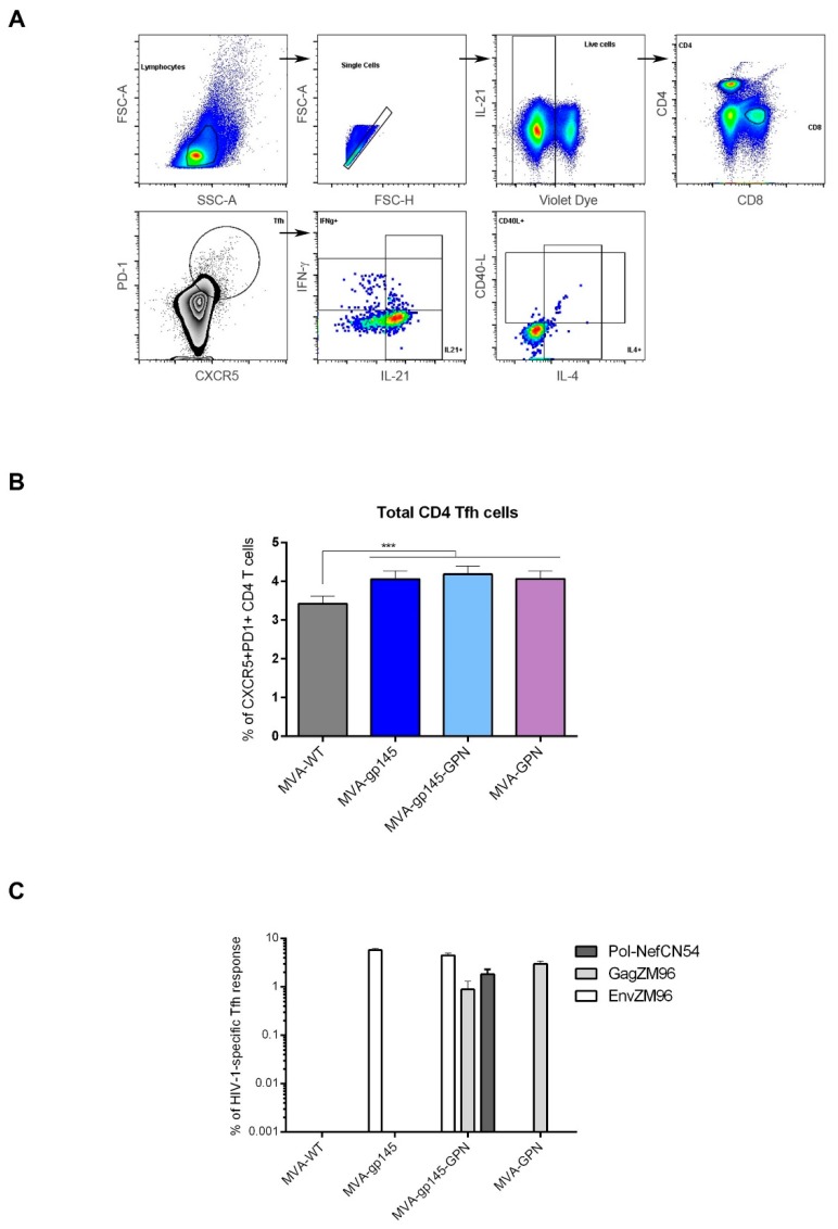
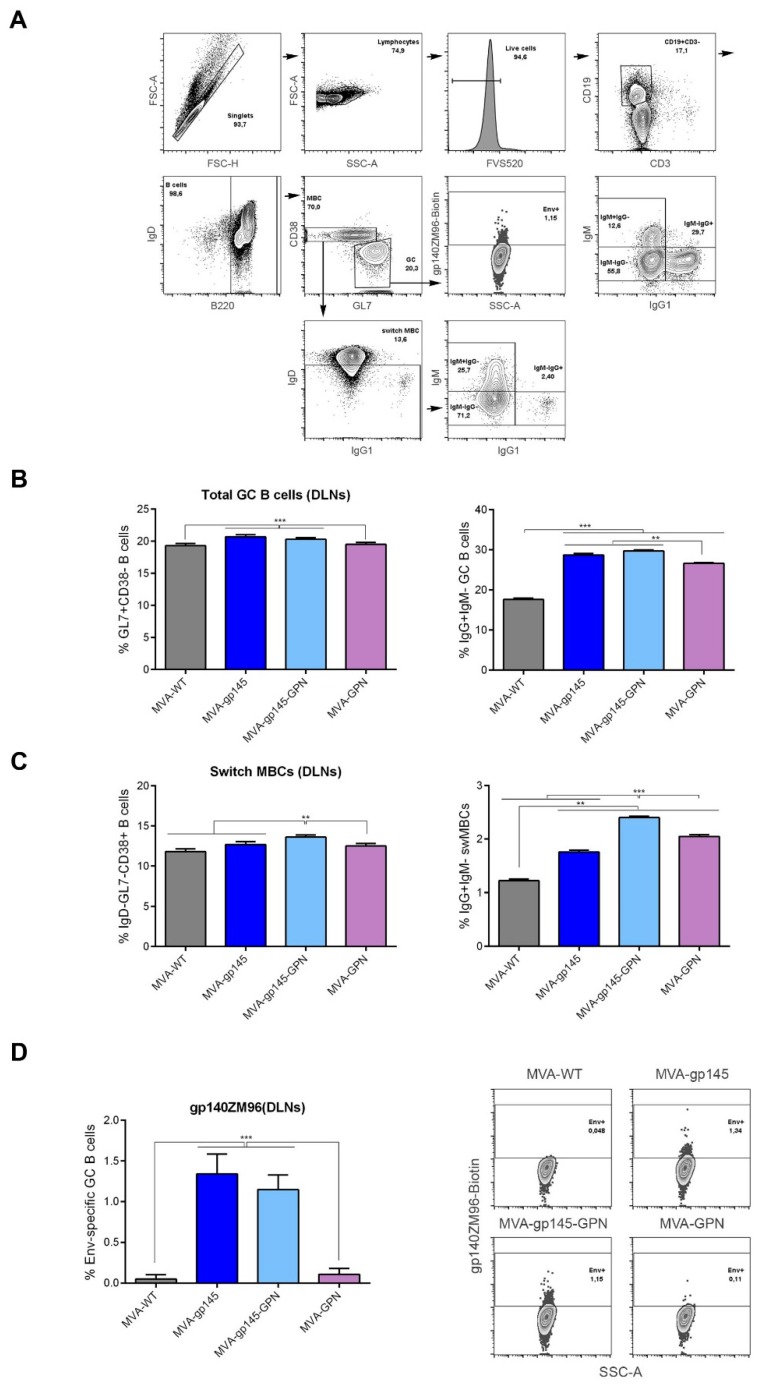
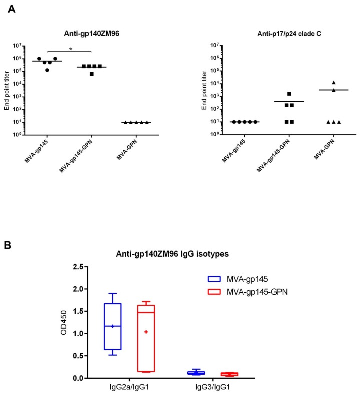
The development of an effective Human Immunodeficiency Virus (HIV) vaccine that is able to stimulate both the humoral and cellular HIV-1-specific immune responses remains a major priority challenge. In this study, we described the generation and preclinical evaluation of single and double modified vaccinia virus Ankara (MVA)-based candidates expressing the HIV-1 clade C membrane-bound gp145(ZM96) trimeric protein and/or the Gag(ZM96)-Pol-Nef(CN54) (GPN) polyprotein that was processed to form Gag-induced virus-like particles (VLPs). In vitro characterization of MVA recombinants revealed the stable integration of HIV-1 genes without affecting its replication capacity. In cells that were infected with Env-expressing viruses, the gp145 protein was inserted into the plasma membrane exposing critical epitopes that were recognized by broadly neutralizing antibodies (bNAbs), whereas Gag-induced VLPs were released from cells that were infected with GPN-expressing viruses. VLP particles as well as purified MVA virions contain Env and Gag visualized by immunoelectron microscopy and western-blot of fractions that were obtained after detergent treatments of purified virus particles. In BALB/c mice, homologous MVA-gp145-GPN prime/boost regimen induced broad and polyfunctional Env- and Gag-specific CD4 T cells and antigen-specific T follicular helper (Tfh) and Germinal Center (GC) B cells, which correlated with robust HIV-1-specific humoral responses. Overall, these results support the consideration of MVA-gp145-GPN vector as a potential vaccine candidate against HIV-1.

Keywords: CD4 T cells; Env-gp145; GC B cells; Gag-Pol-Nef; HIV-1; MVA vaccine; Tfh; VLPs; humoral responses; immunogenicity.


Conflict of interest statement

The authors declare no conflict of interest.

Figures

Figure 1
Figure 1
In vitro characterization of modified vaccinia virus Ankara (MVA)-based recombinant viruses expressing gp145 and/or Gag(ZM96)-Pol-Nef(CN54) (GPN) antigens. (A) Confirmation of HIV-1 gp145 and/or GPN insertion by PCR analysis. Viral DNA was extracted from DF-1 cells infected with MVA-WT, MVA-gp145, MVA-GPN or MVA-gp145-GPN at 5 pfu/cell. Primers thymidine kinase-L (TK-L) and TK-R spanning J2R (TK) flanking sequences or primers HA-MVA and HA-II spanning A56R (haemagglutinin (HA)) flanking sequences were used for PCR analysis of TK or HA loci, respectively. In parental MVA, a 853 bp-product corresponding to parental TK locus is obtained, while in MVA-gp145 and MVA-gp145-GPN a unique 2577 bp-product is observed and in MVA-GPN a unique band of 4500 bp is detected. Regarding the HA locus, a 750 bp-product is observed in MVA-WT, MVA-gp145 and MVA-GPN samples, while in MVA-gp145-GPN a unique 4500 bp-product is obtained. (B) Analysis of virus growth in a permissive cell line. Monolayers of CEF cells were infected with MVA-WT, MVA-gp145, MVA-GPN or MVA-gp145-GPN at 0.1 pfu/cell. At different times post-infection (0, 24, 48 and 72 h), the cells were collected and infectious viruses were quantified by immunostaining plaque assay in DF-1 cells. (C) Time-course expression of HIV-1 antigens by Western-blot analysis. Monolayers of DF-1 (left panels) or HeLa (right panels) cells were infected at 5 pfu/cell with MVA-WT, MVA-gp145, MVA-GPN or MVA-gp145-GPN viruses. At different times post-infection (0, 4, 8 and 24 h), the infected cells were collected, cells extracts fractionated by 10% SDS-PAGE, and analyzed by Western-blot using rabbit polyclonal anti-gp120 (upper panels) or anti-gag p24 (lower panels) antibodies to evaluate the expression of gp145 and Gag antigens, respectively. (D) Fractionation of viral proteins and localization of HIV-1 Env and Gag proteins within purified MVA-based recombinant viruses. Sucrose-purified virions from MVA-based recombinants were sequentially disrupted by treatment with detergents and different fractions were isolated, as described under Materials and Methods. The unfractionated lysate virions (total extract, ET) and the collected fractions (E1, E2, E3 and C) were analysed by Western-blot using anti-gp120 or anti-Gag antibodies.
Figure 2
Figure 2
(AD) Detection of Gag-induced virus-like particles (VLPs) by electron microscopy. HeLa cells were infected with MVA-GPN (A,C) or MVA-gp145-GPN (B,D) recombinant viruses at 5 pfu/cell and at 24 h.p.i. the supernatants were harvested, clarified, purified by ultracentrifugation through a 20% sucrose cushion, followed by a 20%–60% sucrose gradient, and then processed for negative staining (A,B) or immnogold labelling using anti-gp120 antibody (C,D), as described in Materials and Methods. Bar: 100 nm. (E) bNAbs binding profile to membrane-bound gp145 trimeric protein by flow cytometry. HeLa cells infected with MVA-gp145, MVA-gp145-GPN or MVA-GPN viruses were processed for flow cytometry, as described under Materials and Methods using 10 µg/mL of each primary human IgG anti-Env bNAb. The selected panel of human bNAbs targets quaternary V1/V2 N-glycans (PG9, PGT145 and PG16), V3 N-glycans (10–1074 and PGT121), outer domain (OD)-glycans (2G12), CD4 binding site (VRC01, VRC03 and b12) or gp120/gp41ECTO interface (PGT151 and 35O22) on the native Env protein. Samples were acquired in a flow cytometer and geometric Mean Fluorescence Intensity (gMFI) values on the “live cells” gate were used to analyse the results. All of the bNAbs assayed, except PGT151, specifically recognized the gp145 protein expressed by either the single MVA-gp145 or the double MVA-gp145-GPN viruses, although with different affinities. bNAbs targeting V3 N-glycans, OD N-glycans or CD4 binding site exhibited higher gMFIs values than bNAbs recognizing quaternary epitopes either at V2 appex or at gp120-gp41 interface.
Figure 3
Figure 3
Polyfunctional HIV-1-specific CD4 and CD8 T cell immune responses elicited in spleen after prime/boost homologous immunization of mice with different MVA-based recombinant viruses expressing gp145 and/or GPN antigens. (A) Immunization schedule. Groups of 6–8-week-old female mice (n = 5) received the indicated doses of MVA-based recombinant viruses by bilateral intramuscular route (i.m.); three weeks later, the animals were immunized with MVA constructions as in the prime and 10 days post-boost, mice were sacrificed, and the spleens and draining lymph nodes (DLNs) were processed for Intracellular Cytokine Staining (ICS) assay and sera harvested for Enzyme-Linked Immunosorbent Assay (ELISA) to measure the cellular and humoral adaptive immune responses against HIV-1 or VACV antigens, respectively. (B) Magnitude of the HIV-1-specific CD4 (left) or CD8 (right) T cells at 10 days post-boost by ICS assay after the stimulation of splenocytes with the different HIV-1 clade C peptide pools. The total value of each group represents the sum of the percentages of HIV-1-specific CD4 or CD8 T cells secreting CD107a and/or IFN-γ and/or IL-2 and/or TNF-α against different HIV-1 peptide pools. Data are background-subtracted. ***, p < 0.001. (C) Polyfunctional profile of the overall CD4+ T cell response in the different immunization groups. The thirteen positive combinations of the responses are indicated on the x axis, while the percentages of the functionally different cell populations within the total CD4 T cells are represented on the y axis. Non-specific responses that were obtained in the control group were subtracted in all populations. Specific responses are grouped and colour-coded based on the number of functions. C: CD107a; I: IFN-γ; 2: IL-2; T: TNF-α. ***, p < 0.001. (D) Representative phenotypic profiles of the vaccine-induced memory CD4 T cell responses against Env(ZM96) or Gag(ZM96) peptide pools in the indicated immunization groups. The red dots indicate antigen-specific vaccine-induced CD4+ T cells overlaid on the total CD4 T cell subsets (grey).
Figure 4
Figure 4
HIV-1-specific Tfh cell immune response elicited in spleen after prime/boost homologous immunization of mice with different MVA-based recombinant viruses expressing gp145 and/or GPN antigens. (A) Flow cytometry strategy for the identification of total (B) or HIV-1-specific (C) Tfh cells in splenocytes from immunized mice. Singlets were gated on lymphocytes followed by selection of live cells. CD4+CD8 cells were then gated and analyzed based on the expression of the CXCR5 and PD-1 markers. The double positive CXCR5+PD-1+ population was used to identify total Tfh cells. HIV-1-specific Tfh cells were determined by the percentage of CXCR5+PD-1+ cells that produce IFN-γ and/or IL-4 and/or CD40L. FSC-A: forward-scatter area; FSC-H: forward-scatter height; SSC-A: side scatter area. (B) Magnitude of the total CD4 T cells with Tfh phenotype (CXCR5+PD1+) in spleen measured 10 days after the last immunization by ICS assay in non-stimulated (RPMI) lymphocytes derived from immunized animals. All the data are background-subtracted. ***, p < 0.001. (C) Magnitude of the HIV-1-specific Tfh cells in spleen. The total value in each group represents the sum of the percentages of Tfh+ T cells producing IFN-γ and/or IL-4 against HIV-1 peptide pools. All data are background-subtracted.
Figure 5
Figure 5
Env-specific B cell immune response elicited in DLNs after prime/boost homologous immunization of mice with different MVA-based recombinant viruses expressing gp145 and/or GPN antigens. (A) Flow cytometry strategy for the identification of total B cells (B), memory B cells (MBCs) (C) and Env-specific B cells (D) in lymphocytes from DLNs of immunized mice. B cells lymphocytes were identified as B220+ cells gated on singlets/live/CD3CD19+ population. On B cells, we defined GC B cells (GL7+CD38), memory B cells (MBCs) (GL7CD38+) and class-switched MBCs (IgD and IgG1+IgM) populations. Biotinylated gp140 protein was used to identify the frequency of Env-specific GC B cells. FSC-A: forward-scatter area; FSC-H: forward-scatter height; SSC-A: side scatter area. (B,C) Magnitude of the total GC B cells (B) or MBCs (C) in DLNs measured at 10 days post-boost by ICS assay in lymphocytes derived from immunized animals. **, p < 0.005; ***, p < 0.001. (D) Magnitude (left panel) and flow cytometry profiles (right panel) of the Env-specific GC B cells in DLNs measured at 10 days post-boost by ICS assay following the incubation of lymphocytes with biotinylated gp140(ZM96) protein. All data are background-subtracted. ***, p < 0.001.
Figure 6
Figure 6
HIV-1-specific humoral immune response elicited in serum after prime/boost homologous immunization of mice with different MVA-based recombinant viruses expressing gp145 and/or GPN antigens. (A) Levels of anti-gp140- (left panel) or anti-p17/p24- (right panel) specific total IgG binding antibodies measured in individual sera from immunized mice at 10 days post-boost by ELISA. End point titer is defined as the last serum dilution that gave three times the mean OD450 value of the control group. The solid line represents the mean value of each group. *, p < 0.05. (B) Anti-gp140 IgG2a/IgG1 or IgG3/IgG1 ratios elicited in serum from immunized individual mice at 10 days post-boost measured as OD450 at a serum dilution of 1:64000 by ELISA. The “+” symbol indicates the mean value of each group; the box shows the 5th to 95th percentiles and the line in the box indicates the median value of each group.
Figure 7
Figure 7
Polyfunctional VACV E3-specific CD8 T cell immune response elicited in the spleen after prime/boost homologous immunization of mice with different MVA-based recombinant viruses expressing gp145 and/or GPN antigens. (A) Magnitude of the VACV E3-specific CD8 T cells at 10 days post-boost by ICS assay after the stimulation of splenocytes with the VACV E3 peptide. The total value of each group represents the sum of the percentages of HIV-1-specific CD8 T cells secreting CD107a and/or IFN-γ and/or IL-2 and/or TNF-α against VACV E3 peptide. Data are background-subtracted. ***, p < 0.001. (B) Polyfunctional profile of the E3-specific CD8 T cell response in the different immunization groups. Distinct response combinations are indicated on the x axis, while the percentages of the functionally different cell populations within the total CD8 T cells are represented on the y axis. Specific responses are grouped and colour-coded based on the number of functions. C: CD107a; I: IFN-α; 2: IL-2; T: TNF-α. ***, p < 0.001. (C) Flow cytometry profiles of the vaccine-induced memory CD8 T cell phenotype against VACV E3 peptide in the indicated immunization groups.

Similar articles

Cited by

References

    1. Garcia F., de Quiros J.C., Gomez C.E., Perdiguero B., Najera J.L., Jimenez V., Garcia-Arriaza J., Guardo A.C., Perez I., Diaz-Brito V., et al. Safety and immunogenicity of a modified pox vector-based HIV/AIDS vaccine candidate expressing Env, Gag, Pol and Nef proteins of HIV-1 subtype b (MVA-B) in healthy HIV-1-uninfected volunteers: A phase I clinical trial (RISVAC02) Vaccine. 2011;29:8309–8316. doi: 10.1016/j.vaccine.2011.08.098. - DOI - PubMed
    1. Gomez C.E., Najera J.L., Perdiguero B., Garcia-Arriaza J., Sorzano C.O., Jimenez V., Gonzalez-Sanz R., Jimenez J.L., Munoz-Fernandez M.A., de Quiros J.C., et al. The HIV/AIDS vaccine candidate MVA-B administered as a single immunogen in humans triggers robust, polyfunctional, and selective effector memory T cell responses to HIV-1 antigens. J. Virol. 2011;85:11468–11478. doi: 10.1128/JVI.05165-11. - DOI - PMC - PubMed
    1. Gomez C.E., Perdiguero B., Garcia-Arriaza J., Esteban M. Poxvirus vectors as HIV/AIDS vaccines in humans. Hum. Vaccin. Immunother. 2012;8:1192–1207. doi: 10.4161/hv.20778. - DOI - PMC - PubMed
    1. Guardo A.C., Gomez C.E., Diaz-Brito V., Pich J., Arnaiz J.A., Perdiguero B., Garcia-Arriaza J., Gonzalez N., Sorzano C.O.S., Jimenez L., et al. Correction: Safety and vaccine-induced HIV-1 immune responses in healthy volunteers following a late MVA-B boost 4 years after the last immunization. PLoS ONE. 2018;13:e0195915. doi: 10.1371/journal.pone.0195915. - DOI - PMC - PubMed
    1. Joseph S., Quinn K., Greenwood A., Cope A.V., McKay P.F., Hayes P.J., Kopycinski J.T., Gilmour J., Miller A.N., Geldmacher C., et al. A comparative phase I study of combination, homologous subtype-C DNA, MVA, and Env gp140 protein/adjuvant HIV vaccines in two immunization regimes. Front. Immunol. 2017;8:149. doi: 10.3389/fimmu.2017.00149. - DOI - PMC - PubMed

Publication types

MeSH terms

Substances