. 2019 Feb 16;11(2):163.
doi: 10.3390/v11020163.

Pathogenicity and Transmissibility of North American H7 Low Pathogenic Avian Influenza Viruses in Chickens and Turkeys

Affiliations

Pathogenicity and Transmissibility of North American H7 Low Pathogenic Avian Influenza Viruses in Chickens and Turkeys

Ishita Roy Chowdhury et al. Viruses. .

Abstract

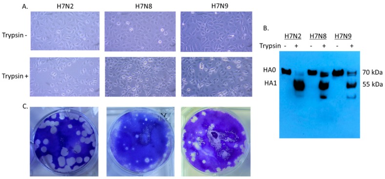
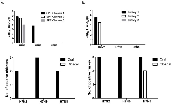
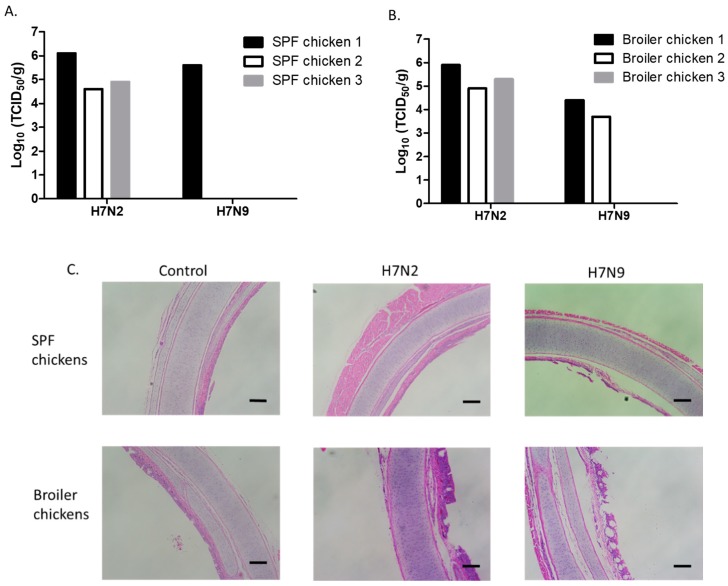
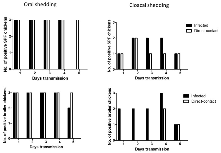
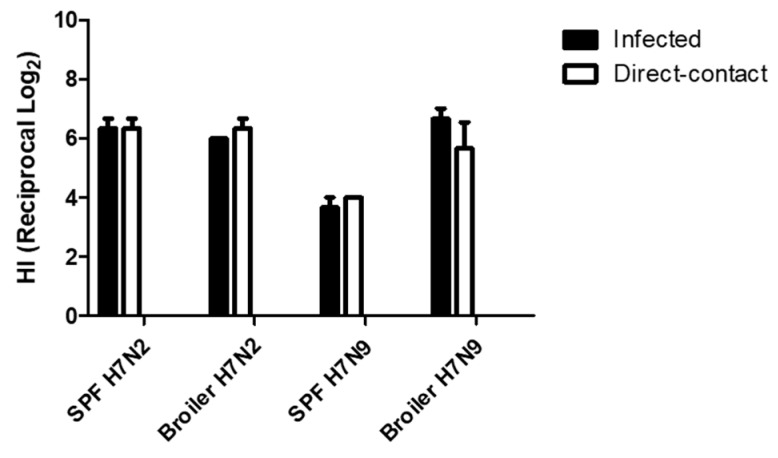
Low pathogenic avian influenza (LPAI) viruses can silently circulate in poultry and wild aquatic birds and potentially mutate into highly pathogenic avian influenza (HPAI) viruses. In the U.S., recent emergence and spread of H7N8 and H7N9 HPAI viruses not only caused devastating losses to domestic poultry but also underscored the capability of LPAI viruses to mutate into HPAI viruses. Therefore, in this study, we evaluated pathogenicity and transmissibility of H7N8 and H7N9 LPAI viruses (the progenitors of HPAI viruses) in chickens and turkeys. We also included H7N2 isolated from an outbreak of LPAI in commercial chickens. H7 viruses replicated more efficiently in the respiratory tract than in the gastrointestinal tract, suggesting that their replication is restricted to the upper respiratory tract. Specifically, H7N2 replicated most efficiently in two-week-old chickens and turkeys. In contrast, H7N8 replicated least efficiently in those birds. Further, replication of H7N2 and H7N9 was restricted in the upper respiratory tract of four-week-old specific-pathogen-free (SPF) and broiler chickens. Despite their restricted replication, the two viruses efficiently transmitted from infected to naïve birds by direct contact, leading to seroconversion of contacted chickens. Our findings suggest the importance of continuous monitoring and surveillance of LPAI viruses in the fields.

Keywords: H7; chickens; low pathogenic avian influenza virus; replication; transmissibility.


Conflict of interest statement

The authors declare no conflict of interest.

Figures

Figure 1
Figure 1
Infectivity of H7N2, H7N8, and H7N9 in MDCK (Madin–Darby canine kidney) cells. MDCK cells were individually infected with H7N2, H7N8, and H7N9 (MOI of 0.1 PFU/cell) in the presence or absence of trypsin. After 12 hpi, the infected cells were visualized directly by photomicroscopy, (20X) (A). Collected cell lysates were subjected to Western blot analysis (B). Plaque formation of H7 viruses in MDCK cells was conducted by plaque assay (C).
Figure 2
Figure 2
Replication and shedding of H7 LPAI viruses in the trachea of 2-week-old SPF (specific-pathogen-free) chickens (A) and turkeys (B). Birds were intranasally inoculated with 200 µL of each virus (107 pfu/mL). Trachea samples were collected for virus titration in MDCK cells on days 3 pi. Virus titers were expressed as 50% tissue culture infectious dose (TCID50/g). Swab (oral and cloacal) samples were collected for virus shedding. Viral replication was confirmed by amplifying the virus in MDCK cells. At 72 h post-infection, viral replication was determined by HA assay.
Figure 3
Figure 3
Replication of H7N2 and H7N9 in the trachea of 4-week-old SPF and broiler chickens. Birds were intranasally inoculated with 200 µL of each virus (107 pfu/mL). Trachea samples were collected from SPF (A) and broiler (B) chickens for virus titration in MDCK cells on days 3 pi. Virus titers were expressed as 50% tissue culture infectious dose (TCID50/g). For histopathology, trachea samples were collected from uninfected or infected 4-week-old SPF and broiler chickens (C). The samples were fixed in phosphate-buffered formalin, were processed for section, and stained with hematoxylin and eosin (scale bar = 2 mm).
Figure 4
Figure 4
Transmission of H7N2 in SPF and broiler chickens. Three infected birds with each virus (107 pfu/mL) and three contact exposure birds (naïve birds) were placed in an isolator in one day apart. From following day, swab samples were collected daily up to 5 days for virus shedding.
Figure 5
Figure 5
Transmission of H7N9 in SPF and broiler chickens. Three infected birds with each virus (107 pfu/mL) and three contact exposure birds (naïve birds) were placed in an isolator in one day apart. From following day, swab samples were collected daily up to 5 days for virus shedding.
Figure 6
Figure 6
Seroconversion of SPF and broiler chickens. Serum samples were collected from Infected and contact exposure birds at 14 days post-exposure. Seroconversion was determined by HI assay.

References

    1. Palese P., Shaw M.L. Orthomyxoviridae: The viruses and their replication. In: Knipe D.M., Howley P.M., editors. Fields Virology. 5th ed. Lippincott Williams & Wilkins; Philadelphia, PA, USA: 2007. pp. 1647–1689.
    1. Sonnberg S., Webby R.J., Webster R.G. Natural history of highly pathogenic avian influenza H5N1. Virus Res. 2013;178:63–77. doi: 10.1016/j.virusres.2013.05.009. - DOI - PMC - PubMed
    1. Alexander D.J. Summary of avian influenza activity in Europe, Asia, Africa, and Australasia, 2002–2006. Avian Dis. 2007;51:161–166. doi: 10.1637/7602-041306R.1. - DOI - PubMed
    1. Webster R.G., Kawaoka Y., Bean W.J. What is the potential of avirulent influenza viruses to complement a cleavable hemagglutinin and generate virulent strains. Virology. 1989;171:484–492. doi: 10.1016/0042-6822(89)90618-1. - DOI - PubMed
    1. Webster R.G., Govorkova E.A. H5N1 influenza—Continuing evolution and spread. N. Engl. J. Med. 2006;355:2174–2177. doi: 10.1056/NEJMp068205. - DOI - PubMed

Publication types

MeSH terms