Review
. 2019 Feb 17;20(4):865.
doi: 10.3390/ijms20040865.

Silver Nanoparticles: Synthesis and Application for Nanomedicine

Affiliations
Review

Silver Nanoparticles: Synthesis and Application for Nanomedicine

Sang Hun Lee et al. Int J Mol Sci. .

Abstract

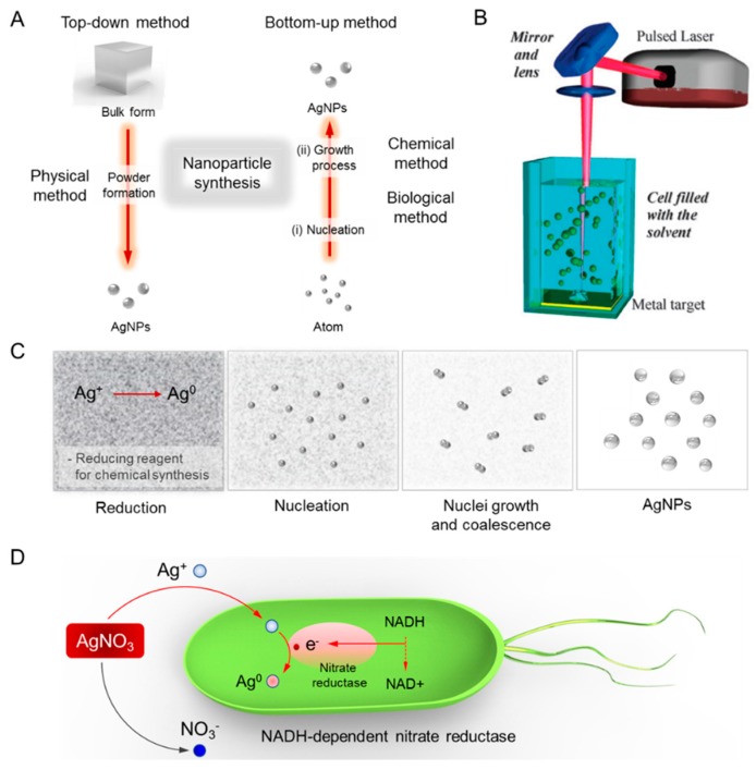
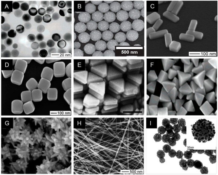
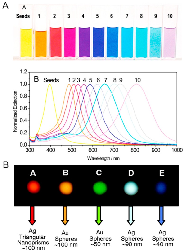
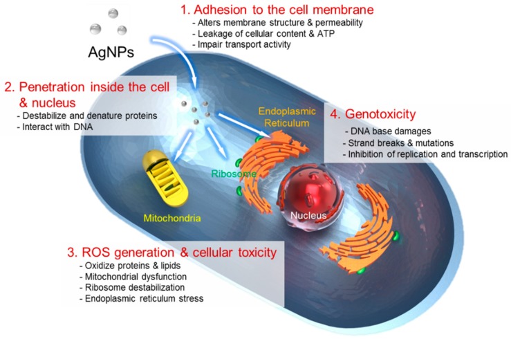
Over the past few decades, metal nanoparticles less than 100 nm in diameter have made a substantial impact across diverse biomedical applications, such as diagnostic and medical devices, for personalized healthcare practice. In particular, silver nanoparticles (AgNPs) have great potential in a broad range of applications as antimicrobial agents, biomedical device coatings, drug-delivery carriers, imaging probes, and diagnostic and optoelectronic platforms, since they have discrete physical and optical properties and biochemical functionality tailored by diverse size- and shape-controlled AgNPs. In this review, we aimed to present major routes of synthesis of AgNPs, including physical, chemical, and biological synthesis processes, along with discrete physiochemical characteristics of AgNPs. We also discuss the underlying intricate molecular mechanisms behind their plasmonic properties on mono/bimetallic structures, potential cellular/microbial cytotoxicity, and optoelectronic property. Lastly, we conclude this review with a summary of current applications of AgNPs in nanoscience and nanomedicine and discuss their future perspectives in these areas.

Keywords: characterization; cytotoxicity; diagnostics; mechanism; nanomedicine; optoelectronics; silver nanomaterial; synthesis.


Conflict of interest statement

The authors declare no conflict of interest.

Figures

Figure 1
Figure 1
Diverse synthesis routes of silver nanoparticles (AgNPs). (A) Top-down and bottom-up methods. (B) Physical synthesis method. Reprinted with permission from [21]. Copyright 2009 Royal Chemical Society. (C) Chemical synthesis method. (D) Plausible synthesis mechanisms of green chemistry. The bioreduction is initiated by the electron transfer through nicotinamide adenine dinucleotide (NADH)-dependent reductase as an electron carrier to form NAD+. The resulting electrons are obtained by Ag+ ions which are reduced to elemental AgNPs.
Figure 2
Figure 2
Representative images of electron microscopy of synthesized Ag nanostructures, demonstrating that diverse sizes and morphologies are made possible by controlling the reaction chemistry. (A) Silver nanosphere [58], (B) Silver necklaces [59], (C) Silver nanobars [60], (D) Silver nanocubes [7], (E) Silver nanoprism [61], (F) Silver bipyramids [62], (G) Silver nanostar [63], (H) Silver nanowire [58], (I) Silver nanoparticle embedded silica particle [64]. All figures were reprinted with permission from the publisher of each article.
Figure 3
Figure 3
(A) Photograph of silver nanoprisms (top) and corresponding optical spectra changes of nanoprisms (bottom). Control on the edge-length of nanoprisms allows the plasmon resonance to be tuned across the visible and near-infrared portions of the spectrum. Reprinted with permission from [55]. Copyright 2008 Wiley-VCH. (B) Dark field microscopy images of (left to right) 100 nm diameter silver triangular nanoprism, 90 nm diameter silver nanosphere, and 40 nm diameter silver nanosphere, illustrating the ability to tune the scattering color of silver nanoparticle labels based on size and shape. Reprinted with permission from [56]. Copyright 2001 American Association for the Advancement of Science.
Figure 4
Figure 4
The four main routes of cytotoxic mechanism of AgNPs. 1, AgNPs adhere to the surface of a cell, damaging its membrane and altering the transport activity; 2, AgNPs and Ag ions penetrate inside the cell and interact with numerous cellular organelles and biomolecules, which can affect corresponding cellular function; 3, AgNPs and Ag ions participate in the generation of reactive oxygen species (ROS) inside the cell leading to a cell damage and; 4, AgNPs and Ag ions induce the genotoxicity.
Figure 5
Figure 5
Controlled overgrowth of Ag for bimetallic nanocrystals. (A) Schematic illustration showing the site-selective growth of Ag on each cubic seed and corresponding transmission electron microscopy (TEM) images. Well-controlled bimetallic nanocrystals were fabricated along the directed size and number of facets on a cubic Pd seed. The white dashed lines in the TEM indicate the position of the cubic Pd seed. (B) Extinction spectra of the Pd–Ag bimetallic nanocrystals with Ag growing on different numbers of faces of the cubic Pd seed. The LSPR peak blue-shifted with the increase in the number of faces involved in the Ag growth. Reprinted with permission from [80]. Copyright 2012 American Chemical Society.
Figure 6
Figure 6
Plasmonic AgNPs for plasmonic nanoantennas and diagnostics. (A) Single-layer AgNP surface-enhanced Raman scattering (SERS) film for a large-scale hot spot. (i) Scanning electron microscopy (SEM) image of a superlattice of 6 nm. AgNPs were used as a homogeneous single-molecule SERS substrate. Illustration shows an interparticle gap for hot spots, which is regulated by the length of a thiolate chain. (ii) Two Raman spectra of single-layered SERS film (left) and quartz surface (right). The enhancement factor was estimated to be larger than 1.2 × 10⁷. Reprinted with permission from [94]. Copyright 2015 American Chemical Society. (B) Metal-film induced plasmon resonance tuning of AgNPs. (i) Schematic illustration of optical scattering spectra of AgNPs on different substrates. (ii) Single AgNP spectra of AgNPs on a silica spacer layer of varying thickness d (nm) on a glass substrate with a 50 nm gold film. The inset is a dark-field image of AgNPs with the corresponding color. The dotted lines represent single particle spectra of AgNPs on a plain glass substrate. Reprinted with permission from [103]. Copyright 2010 American Chemical Society. (C) SERS-based intracellular imaging using alkyne-AgNPs nanoprobes. (i) The structure of colloidal alkyne-AgNP clusters with nano-sized interparticle gaps. (ii) Extinction spectra of the alkyne-AgNPs nanoprobe. The resonance peaks at 400 nm shifted around 520 nm after metal functionalization. (iii) Computational simulation of the far- and near-field optical responses. Intensity distributions of the single particle mode (upper-panels) and the dimer mode (bottom-panels) (iv) Intracellular Raman imaging of a AgNP nanoprobe within the cytoplasmic space of fibroblast. Distinguishable hot spots were highlighted by color-dots related to Raman intensity of the akyne 2045 cm−1 band. Reprinted with permission from [104]. Copyright 2018 Nature Publishing Group. (D) Multiplexed detection with a tunable wavelength of AgNPs. (i) Different colors of AgNPs during a stepwise growth. (ii) Corresponding absorption spectra with varying sizes of AgNPs, such as 30, 41, and 47 nm. (iii) Individual testing of Yellow Fever virus (YFV) NS1 protein, Zaire Ebola virus (ZEBOV) glycoprotein (GP), and Dengue virus (DENV) NS protein using AgNPs. Orange, red, and green AgNPs were conjugated with monoclonal antibodies specific to YFV NS1, ZEBOV GP, and DENV NS, respectively. (iv) Multiplexed detection using different AgNPs-based lateral flow assays. Reprinted with permission from [105]. Copyright 2015 Royal Society of Chemistry.
Figure 7
Figure 7
Surface-enhanced fluorescence. (A) Metal-enhanced fluorescence on a Ag film. (i) The photograph shows fluorescence spots on quartz (top) and silver (bottom) taken through 530 nm long pass filter for Cy3-DNA. (ii) Emission spectra of Cy3-DNA on APS-treated slides, with (solid line) and without silver island films (dotted line). Reprinted with permission from [113]. Copyright 2003 Future Science Group. (B) Schematic illustration of an aptamer-based AgNP nanosensor, showing the ‘off’ state via fluorophore quenching within short distances (left) and ‘on’ state via turn-on fluorescence signal (right) based on the spacing distance between the Cyanine 3 and the AgNP surface in the detection of adenosine. Reprinted with permission from [114]. Copyright 2012 Elsevier.

References

    1. Millstone J.E., Hurst S.J., Métraux G.S., Cutler J.I., Mirkin C.A. Colloidal gold and silver triangular nanoprisms. Small. 2009;5:646–664. doi: 10.1002/smll.200801480. - DOI - PubMed
    1. Lee S.H., Rho W.-Y., Park S.J., Kim J., Kwon O.S., Jun B.-H. Multifunctional self-assembled monolayers via microcontact printing and degas-driven flow guided patterning. Sci. Rep. 2018;8:16763. - PMC - PubMed
    1. Lee S.H., Sung J.H., Park T.H. Nanomaterial-based biosensor as an emerging tool for biomedical applications. Ann. Biomed. Eng. 2012;40:1384–1397. doi: 10.1007/s10439-011-0457-4. - DOI - PubMed
    1. Syafiuddin A., Salmiati, Salim M.R., Kueh A.B.H., Hadibarata T., Nur H. A Review of silver nanoparticles: Research trends, global consumption, synthesis, properties, and future Challenges. J. Clin. Chem. Soc. 2017;64:732–756. doi: 10.1002/jccs.201700067. - DOI
    1. Kumar A., Vemula P.K., Ajayan P.M., John G. Silver-nanoparticle-embedded antimicrobial paints based on vegetable oil. Nat. Mater. 2008;7:236–241. doi: 10.1038/nmat2099. - DOI - PubMed

LinkOut - more resources