. 2019 Feb 13;9(2):256.
doi: 10.3390/nano9020256.

Synthesis and Characterization of Elongated-Shaped Silver Nanoparticles as a Biocompatible Anisotropic SERS Probe for Intracellular Imaging: Theoretical Modeling and Experimental Verification

Affiliations

Synthesis and Characterization of Elongated-Shaped Silver Nanoparticles as a Biocompatible Anisotropic SERS Probe for Intracellular Imaging: Theoretical Modeling and Experimental Verification

Carlos Caro et al. Nanomaterials (Basel). .

Abstract

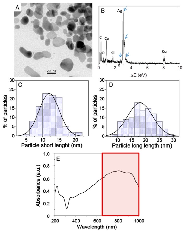
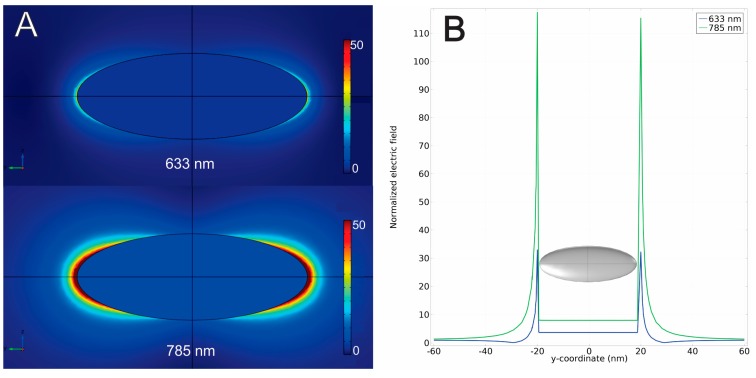
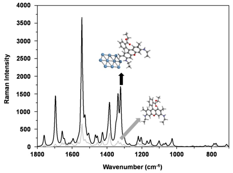
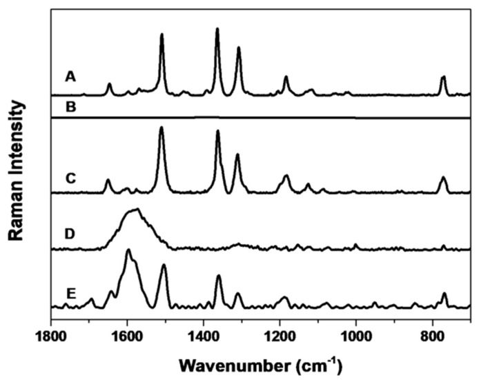
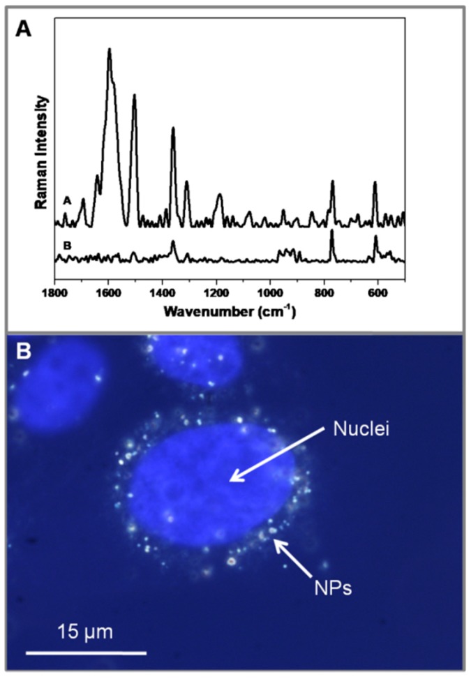
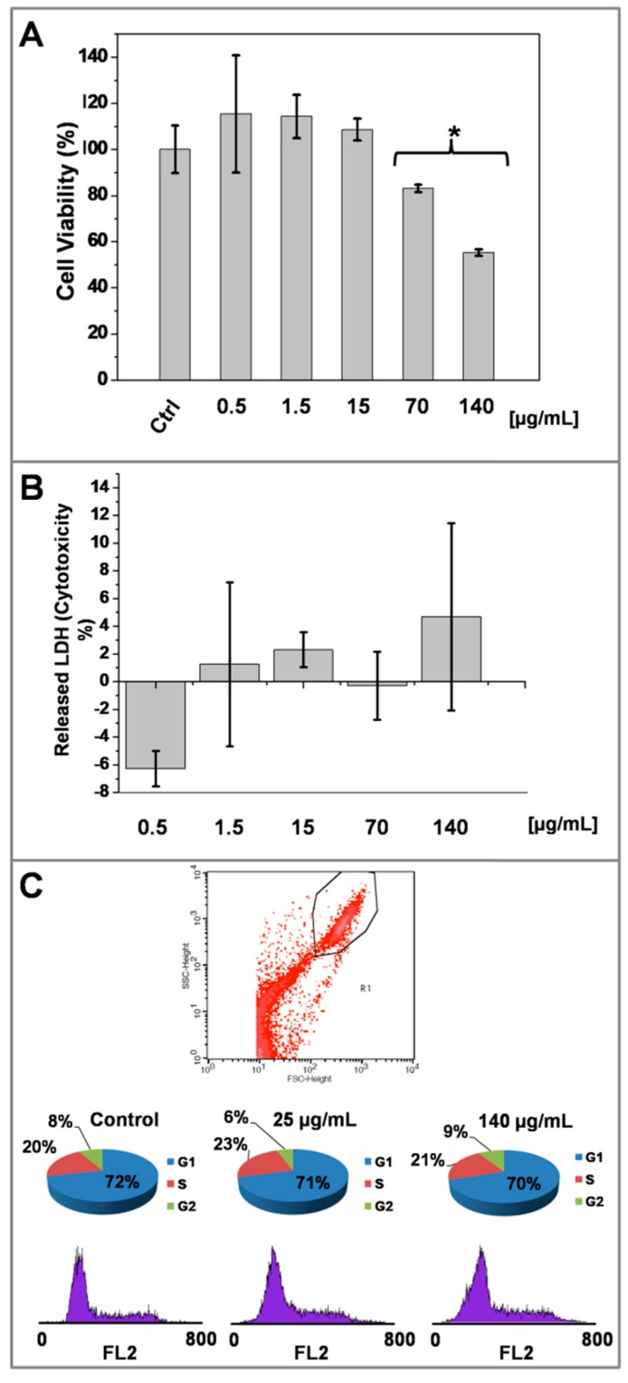
Progress in the field of biocompatible SERS nanoparticles has promising prospects for biomedical applications. In this work, we have developed a biocompatible Raman probe by combining anisotropic silver nanoparticles with the dye rhodamine 6G followed by subsequent coating with bovine serum albumin. This nanosystem presents strong SERS capabilities in the near infrared (NIR) with a very high (2.7 × 10⁷) analytical enhancement factor. Theoretical calculations reveal the effects of the electromagnetic and chemical mechanisms in the observed SERS effect for this nanosystem. Finite element method (FEM) calculations showed a considerable near field enhancement in NIR. Using density functional quantum chemical calculations, the chemical enhancement mechanism of rhodamine 6G by interaction with the nanoparticles was probed, allowing us to calculate spectra that closely reproduce the experimental results. The nanosystem was tested in cell culture experiments, showing cell internalization and also proving to be completely biocompatible, as no cell death was observed. Using a NIR laser, SERS signals could be detected even from inside cells, proving the applicability of this nanosystem as a biocompatible SERS probe.

Keywords: SERS; cancer; cell labeling; density functional theory calculations; finite element method; surface enhanced Raman scattering.


Conflict of interest statement

The authors declare no conflict of interest.

Figures

Figure 1
Figure 1
Microstructural characterization of AgNPs by TEM (A), EDX analysis of AgNPs (B), in which arrows indicate silver peaks. Particles short-length (C) and long-length (D) histograms-based on the measurements of over 150 nanoparticles. UV-Vis spectrum of AgNPs in solution (E) showing a maximum absorption in the NIR region (650–1350 nm in a red box). Signals from the elements Si, Cu and C are originated from carbon-coated copper TEM grid.
Figure 2
Figure 2
Local electric field enhancement (A) in the vicinity of one AgNP for incident radiation at 633 nm (A, top scheme) and at 785 nm (A, bottom scheme). Graphical representation of the electric field strength (B) (normalized to the incident field) along the y-axis of the simulation crossing the points of greater electric field intensity around the apexes of the AgNP.
Figure 3
Figure 3
DFT calculation of R6G alone (grey line) and R6G interacting with a thin layer of 10 atoms of Ag (black line). The Ag layer causes a SERS effect leading to an increase in Raman signal intensity of one order of magnitude.
Figure 4
Figure 4
Raman and SERS spectra from different steps in the nanosystem synthesis. Raman spectra of pure R6G solution (A) and R6G at the same concentration used in the AgNP@R6G@BSA nanosystem (B). No spectrum is obtained from (B) because of the very low concentration of R6G. SERS spectra of AgNP@R6G (C), which is Raman active but has a very limited colloidal stability. AgNP@BSA is not Raman active (D). Raman spectra of AgNP@R6G@BSA nanosystem (E).
Figure 5
Figure 5
SERS spectra of AgNP@R6G@BSA (A. Inset A) and AgNP@R6G@BSA-treated human carcinoma cell line (A431) (B. Inset B). Overlay images of blue fluorescence channel (Hoechst dye labelling cell nuclei) and dark field microscopy (DFM) reveal nanoparticle internalization.
Figure 6
Figure 6
Effect of AgNP@R6G@BSA on A431 human epidermoid carcinoma cell line biocompatibility. Mitochondrial function (MTT reduction) after 24 h of treatment with increasing concentrations of AgNPs (A). Lactate dehydrogenase release assay (LDH) after 24 h of treatment with increasing concentrations of AgNP@R6G@BSA (B). Flow cytometry analysis of cell cycle after 24 h of treatment with increasing concentrations of AgNP@R6G@BSA (C). Data represented the mean ± SD. Measurements were made in three replicas for each experimental condition.

References

    1. Sánchez-Iglesias A., Aldeanueva-Potel P., Ni W., Pérez-Juste J., Pastoriza-Santos I., Alvarez-Puebla R.A., Mbenkum B.N., Liz-Marzán L.M. Chemical seeded growth of Ag nanoparticle arrays and their application as reproducible SERS substrates. Nano Today. 2010;5:21–27. doi: 10.1016/j.nantod.2010.01.002. - DOI
    1. Alvarez-Puebla R.A., Liz-Marzán L.M. SERS-Based Diagnosis and Biodetection. Small. 2010;6:604–610. doi: 10.1002/smll.200901820. - DOI - PubMed
    1. Leopold N., Lendl B. A New Method for Fast Preparation of Highly Surface-Enhanced Raman Scattering (SERS) Active Silver Colloids at Room Temperature by Reduction of Silver Nitrate with Hydroxylamine Hydrochloride. J. Phys. Chem. B. 2003;107:5723–5727. doi: 10.1021/jp027460u. - DOI
    1. Abalde-Cela S., Abell C., Alvarez-Puebla R.A., Liz-Marzán L.M. Real Time Dual-Channel Multiplex SERS Ultradetection. J. Phys. Chem. Lett. 2014;5:73–79. doi: 10.1021/jz402419k. - DOI - PubMed
    1. Piorek B.D., Andreou C., Moskovits M., Meinhart C.D. Discrete Free-Surface Millifluidics for Rapid Capture and Analysis of Airborne Molecules Using Surface-Enhanced Raman Spectroscopy. Anal. Chem. 2014;86:1061–1066. doi: 10.1021/ac402628t. - DOI - PubMed