

The importance of the PD-1/PD-L1 pathway at the maternal-fetal interface
- PMID: 30782114
- PMCID: PMC6381664
- DOI: 10.1186/s12884-019-2218-6
The importance of the PD-1/PD-L1 pathway at the maternal-fetal interface
Abstract
Background: Our goal with this study was to investigate the contribution of PD-1/PD-L1 immune-checkpoint pathway to maternal immunotolerance mechanisms.
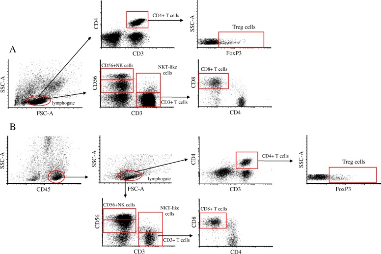
Methods: Thirteen healthy pregnant women and 10 non-pregnant controls were involved in this project. PBMCs and DICs were isolated from peripheral blood and from decidual tissues. After the characterization of different immune cell subsets, we used fluorochrome-conjugated monoclonal antibodies to measure the expression level of PD-1, PD-L1, NKG2D, and CD107a molecules by flow cytometry.
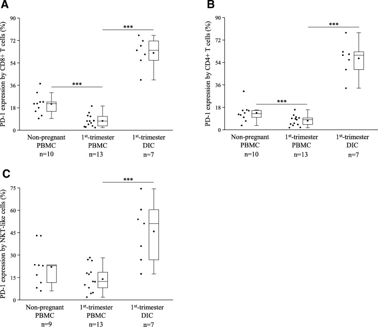
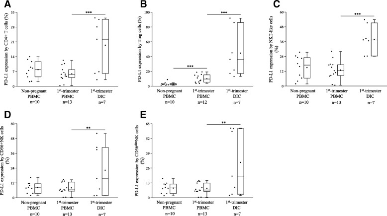
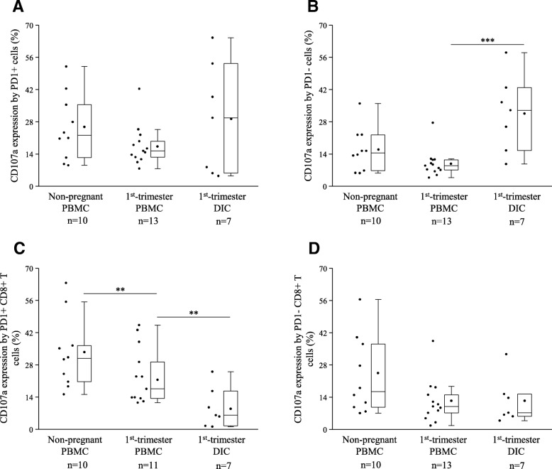
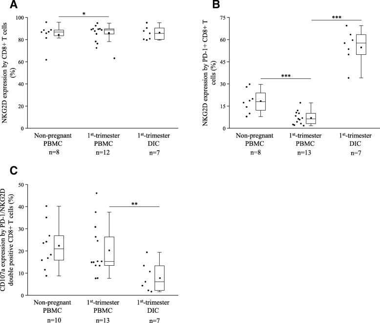
Results: We measured significant alternations in the proportion of decidual immune cell subsets compared to the periphery. Elevated PD-1 expression by decidual CD8+ T, CD4+ T, and NKT-like cells were also detected accompanied by the increased PD-L1 expression by decidual CD4+ T, Treg, NKT-like and CD56 + NK cell subsets compared to peripheral blood. The cytotoxic potential was significantly higher in PD-1- decidual immune cells compared to the periphery, however we measured a significantly lower cytotoxicity in the decidual PD-1+ CD8+ T cells compared with the peripheral subsets. An activation receptor NKG2D expression was decreased by the PD-1+ CD8+ T subsets in the first trimester compared to non-pregnant condition but the expression level of the decidual counterparts was significantly elevated compared to the periphery. The cytotoxic potential of decidual PD1/NKG2D double positive CD8+ T cells was significantly decreased compared to the peripheral subsets.
Conclusions: Based on our results we assume that PD-1/PD-L1 pathway might have a novel role in the maintaining of the local immunological environment. Accompanied by NKG2D activating receptor this checkpoint interaction could regulate decidual CD8 Tc cell subsets and may contribute maternal immunotolerance.
Keywords: Maternal immunotolerance; Maternal-fetal interface; PD-1; PD-L1; Pregnancy.
Conflict of interest statement
Ethics approval and consent to participate
The collection of the samples and experimental procedures were approved by the Regional Ethics Committee at the Medical School, University of Pécs (approved protocol registration number: 6149) and written informed consent was obtained from all participant. The study protocol conforms to the ethical guidelines of the 1975 Declaration of Helsinki.
Consent for publication
Not applicable.
Competing interests
The authors declare that they have no competing interests.
Publisher’s Note
Springer Nature remains neutral with regard to jurisdictional claims in published maps and institutional affiliations.
Figures
Similar articles
-
Analysis of TCR Repertoire and PD-1 Expression in Decidual and Peripheral CD8+ T Cells Reveals Distinct Immune Mechanisms in Miscarriage and Preeclampsia.Front Immunol. 2020 Jun 3;11:1082. doi: 10.3389/fimmu.2020.01082. eCollection 2020. Front Immunol. 2020. PMID: 32582176 Free PMC article.
-
Frequencies of PD-1- and PD-L1- positive T CD3+CD4+, T CD3+CD8+ and B CD19+ lymphocytes and its correlations with other immune cells in patients with recurrent furunculosis.Microb Pathog. 2019 Jan;126:85-91. doi: 10.1016/j.micpath.2018.10.019. Epub 2018 Oct 19. Microb Pathog. 2019. PMID: 30342909
-
The role of the PD-1/PD-L1 axis in macrophage differentiation and function during pregnancy.Hum Reprod. 2019 Jan 1;34(1):25-36. doi: 10.1093/humrep/dey347. Hum Reprod. 2019. PMID: 30500923
-
[Efficacy of PD-1/PD-L1 immune checkpoint inhibitors and PD-L1 testing in thoracic cancers].Ann Pathol. 2017 Feb;37(1):61-78. doi: 10.1016/j.annpat.2016.12.009. Epub 2017 Feb 3. Ann Pathol. 2017. PMID: 28162296 Review. French.
-
PD-1/PD-L1 Pathway in Breast Cancer.Oncol Res Treat. 2017;40(5):294-297. doi: 10.1159/000464353. Epub 2017 Mar 27. Oncol Res Treat. 2017. PMID: 28346916 Review.
Cited by
-
Surface Immune Checkpoints as Potential Biomarkers in Physiological Pregnancy and Recurrent Pregnancy Loss.Int J Mol Sci. 2024 Aug 29;25(17):9378. doi: 10.3390/ijms25179378. Int J Mol Sci. 2024. PMID: 39273326 Free PMC article.
-
Effects of PACAP Deficiency on Immune Dysfunction and Peyer's Patch Integrity in Adult Mice.Int J Mol Sci. 2024 Oct 3;25(19):10676. doi: 10.3390/ijms251910676. Int J Mol Sci. 2024. PMID: 39409005 Free PMC article.
-
Exploring Natural Killer Cell Testing in Embryo Implantation and Reproductive Failure: An Overview of Techniques and Controversies.Reprod Sci. 2024 Mar;31(3):603-632. doi: 10.1007/s43032-023-01372-z. Epub 2023 Oct 18. Reprod Sci. 2024. PMID: 37853155 Review.
-
Placental Immune Tolerance and Organ Transplantation: Underlying Interconnections and Clinical Implications.Front Immunol. 2021 Aug 3;12:705950. doi: 10.3389/fimmu.2021.705950. eCollection 2021. Front Immunol. 2021. PMID: 34413856 Free PMC article. Review.
-
Role of maternal-fetal immune tolerance in the establishment and maintenance of pregnancy.Chin Med J (Engl). 2024 Jun 20;137(12):1399-1406. doi: 10.1097/CM9.0000000000003114. Epub 2024 May 9. Chin Med J (Engl). 2024. PMID: 38724467 Free PMC article. Review.
References
-
- Lin H, Mosmann TR, Guilbert L, Tuntipopipat S, Wegmann TG. Synthesis of T helper 2-type cytokines at the maternal-fetal interface. J Immunol. 1993;151:4562–4573. - PubMed
MeSH terms
Substances
Grants and funding
- NKFIH K119529/National Research, Development and Innovation Office
- KA-2018-07/Általános Orvostudományi Kar, Pécsi Tudományegyetem
- KA-2018-18/Általános Orvostudományi Kar, Pécsi Tudományegyetem
- EFOP-3.6.3-VEKOP-16-2017-00009/Development of scientific workshops of medical, health sciences and pharmaceutical educations
LinkOut - more resources
Full Text Sources
Research Materials
