. 2019 Feb 19;14(1):58.
doi: 10.1186/s13018-019-1082-4.

Burn and thoracic trauma alters fracture healing, systemic inflammation, and leukocyte kinetics in a rat model of polytrauma

Affiliations

Burn and thoracic trauma alters fracture healing, systemic inflammation, and leukocyte kinetics in a rat model of polytrauma

Lauren H Mangum et al. J Orthop Surg Res. .

Abstract

Background: Singular traumatic insults, such as bone fracture, typically initiate an appropriate immune response necessary to restore the host to pre-insult homeostasis with limited damage to self. However, multiple concurrent insults, such as a combination of fracture, blunt force trauma, and burns (polytrauma), are clinically perceived to result in abnormal immune response leading to inadequate healing and resolution. To investigate this phenomenon, we created a model rat model of polytrauma.

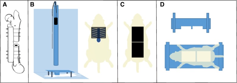
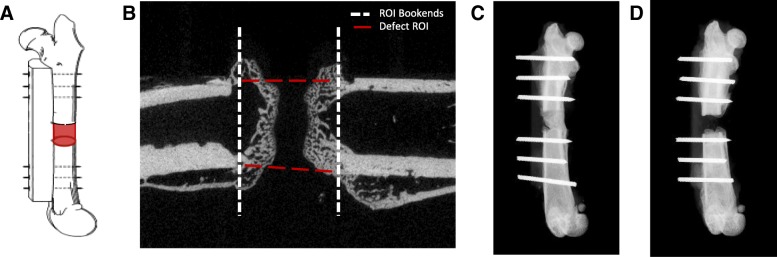
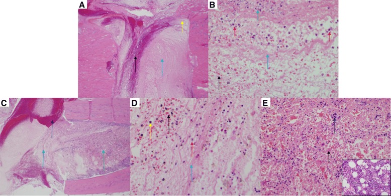
Methods: To investigate relationship between polytrauma and delayed healing, we created a novel model of polytrauma in a rat which encompassed a 3-mm osteotomy, blunt chest trauma, and full-thickness scald burn. Healing outcomes were determined at 5 weeks where the degree of bone formation at the osteotomy site of polytrauma animals was compared to osteotomy only animals (OST).

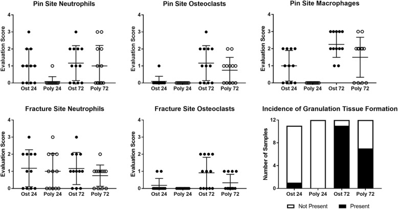
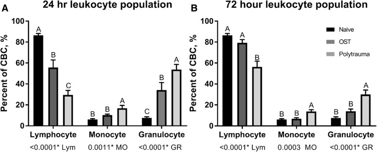
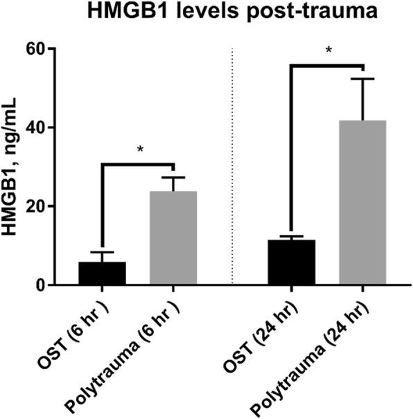
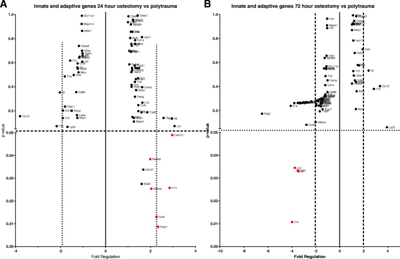
Results: We observed significant differences in the bone volume fraction between polytrauma and OST animals indicating that polytrauma negatively effects wound healing. Polytrauma animals also displayed a significant decrease in their ability to return to pre-injury weight compared to osteotomy animals. Polytrauma animals also exhibited significantly altered gene expression in osteogenic pathways as well as the innate and adaptive immune response. Perturbed inflammation was observed in the polytrauma group compared to the osteotomy group as evidenced by significantly altered white blood cell (WBC) profiles and significantly elevated plasma high-mobility group box 1 protein (HMGB1) at 6 and 24 h post-trauma. Conversely, polytrauma animals exhibited significantly lower concentrations of plasma TNF-alpha (TNF-α) and interleukin 6 (IL-6) at 72 h post-injury compared to OST.

Conclusions: Following polytrauma with burn injury, the local and systemic immune response is divergent from the immune response following a less severe singular injury (osteotomy). This altered immune response that follows was associated with a reduced capacity for wound healing.

Keywords: Extremity fracture; Inflammation; Nonunion; Polytrauma.


Conflict of interest statement

Ethics approval and consent to participate

This study was conducted in compliance with the Animal Welfare Act, the implementing Animal Welfare Regulations, and the principles of the Guide for the Care and Use of Laboratory Animals. All in vivo animal procedures described herein were conducted under appropriate anesthesia with pre- and post-procedural analgesia and monitoring as approved by the USAISR Institutional Animal Care and Use Committee.

Consent for publication

Not applicable

Competing interests

The authors declare that they have no competing interests.

Publisher’s Note

Springer Nature remains neutral with regard to jurisdictional claims in published maps and institutional affiliations.

Figures

Fig. 1
Fig. 1
Representative images of surgical and injury methods. a Osteotomy was achieved via internal fixation with a polyacetal plate and threaded K-wires. b Blunt chest trauma device and placement of animal in apparatus to ensure all energy is directed toward the lungs. c Scalded area on the dorsal side of the animal. d Scald burn plexiglass mold
Fig. 2
Fig. 2
Injured and non-injured lungs and skin. Representative images (H&E and Masson’s Trichrome) from a normal skin (naïve) (H&E; 100x). b Normal skin (naïve) (Masson’s Trichrome; × 100). c Scalded skin (H&E; × 100). Note the coagulated stroma (black arrow), necrotic epidermis (blue arrow), necrotic hair follicle (red arrow), and necrotic sebaceous gland (yellow arrow) compared to panel a. d Scalded skin (Masson’s Trichrome; × 100). Note that the coagulated stroma stains red compared to the normal stroma (black arrow) in panel b. e Normal lung (naïve) (H&E; × 400). f Lung from a rat subjected to blunt force trauma (H&E; × 400). Note the thickening of the alveolar septa with fibrin and inflammatory cells (black arrow) and alveoli which contain hemorrhage (blue arrow) and fibrin (red arrow) and mixed with inflammatory cells (yellow arrow). Note that alveoli normally contain few macrophages (panel e; green arrow), but are not associated with alveolar septal lesions, hemorrhage, or fibrin
Fig. 3
Fig. 3
μCT and radiographic images of OST and polytrauma. a Representation of μCT volume of interest. b ROI bookends encompassed the central 251 slices in between the first distal and proximal slice that did not include cortical bone. c Representative radiograph of the 5-week osteotomy group (n = 8). d Representative radiograph of the 5-week polytrauma group (n = 7)
Fig. 4
Fig. 4
Quantification of immune cell infiltration into pin site and fracture site. Histological analysis was conducted on femurs at 24 and 72 h post-trauma (n = 6/group). Femur sections were cut at a thickness of 8 μm, deparaffinized, and stained with hematoxylin and eosin (H&E) for analysis. The degree of immune cell infiltration and inflammation scored by a veterinary pathologist on a scale of 0–4 and compared between the osteotomy and polytrauma groups within each time point. Significances were determined by Mann-Whitney test, p < 0.05; n = 11–12 animals per time point
Fig. 5
Fig. 5
Osteotomy site in a rat femur (H&E) at 72 h post-trauma. Sections of the osteotomy site (n = 6/group) were stained with hematoxylin and eosin stain. a Representative images of the fracture site from the osteotomy group. There is no callus formation. The osteotomy site is filled with moderate hemorrhage (black arrow) and marked fibrin (blue arrow) (× 40). b Higher magnification (× 600) of the site identified by the yellow arrow in (a). Note the degenerate neutrophils (red arrows), karyorrhectic and cellular debris (green arrow), fibrin (blue arrow), and hemorrhage (black arrow). c Representative image of the fracture site from the polytrauma group. There is no callus formation. The polytrauma osteotomy site is filled with marked hemorrhage (black arrow) and fibrin (blue arrow) (× 40). d Higher magnification (× 600) of the site identified by the blue arrow in c. Note the accumulation of fibrin (blue arrow), hemorrhage (black arrow), few neutrophils (red arrow), and hemosiderin-laden macrophages (orange arrow). The presence of hemosiderin-laden macrophages is indicative of phagocytosis of erythrocytes and hemoglobin. e Higher magnification (× 400) of the bone marrow identified with the green arrow in c. Note the cellular necrosis (black arrows) and marked loss of the cells and adipose tissue compared to the inset (normal bone marrow from sham; × 600)
Fig. 6
Fig. 6
White blood cell concentration following trauma. A comparison in the percentage of total cells population identified as lymphocytes, monocytes, and granulocytes, at two time points: a 24 h and b 72 h in naïve (far left), osteotomy (middle), and polytrauma animals (far right). Values are presented as a percentage of 100 (n = 8/group 24 h post-injury; n = 7, 72 h post osteotomy; n = 6, 72 h post polytrauma; n = 8 naïve). Statistically significant difference between trauma animal cohorts within each cell type, levels not connected by the same letter are significantly different, p < 0.05
Fig. 7
Fig. 7
Systemic concentrations of HMGB1 at 6 and 24 h post-trauma. Plasma HMGB1 levels were measured by ELISA at 6 h (left) (n = 3/OST and n = 5/polytrauma) and 24 h post-trauma (right) (n = 8/OST and 5/polytrauma). At both time points, HMGB1 was significantly increased in the polytrauma group (p < 0.05)
Fig. 8
Fig. 8
Local expression of inflammatory genes at 24 h and 72 h post-trauma. Innate and adaptive responses were compared between osteotomy and polytrauma animals in RNA isolated from the femur following trauma by RT2 PCR Array. a Out of 84 genes, 6 were significantly (p < 0.05) upregulated at least twofold in the polytrauma cohort compared to osteotomy at 24 h and b 4 genes were significantly downregulated at 72 h (N = 5/group)

References

    1. Bastian O, Pillay J, Alblas J, Leenen L, Koenderman L, Blokhuis T. Systemic inflammation and fracture healing. J Leukoc Biol. 2011;89(5):669–673. - PubMed
    1. Hak DJ, Fitzpatrick D, Bishop JA, Marsh JL, Tilp S, Schnettler R, et al. Delayed union and nonunions: epidemiology, clinical issues, and financial aspects. Injury. 2014;45(Supplement 2):S3–S7. - PubMed
    1. Dunlop S, Ekegren C, Edwards E, de Steiger R, Page R, Gabbe B. Hospital admissions and inpatient costs of non-union, delayed union and mal-union following long bone fracture. Value Health. 2016;19(7):A916.
    1. Hoff P, Gaber T, Strehl C, Jakstadt M, Hoff H, Schmidt-Bleek K, et al. A pronounced inflammatory activity characterizes the early fracture healing phase in immunologically restricted patients. Int J Mol Sci. 2017;18(3):583. - PMC - PubMed
    1. Hankenson KD, Zimmerman G, Marcucio R. Biological perspectives of delayed fracture healing. Injury. 2014;45(Supplement 2):S8–S15. - PMC - PubMed

MeSH terms