

Comment
doi: 10.1073/pnas.1900799116.
Epub 2019 Feb 19.
Nucleotide-dependent conformational changes in the actin filament: Subtler than expected
Affiliations
- PMID: 30782816
- PMCID: PMC6410793
- DOI: 10.1073/pnas.1900799116
Item in Clipboard
Comment
Nucleotide-dependent conformational changes in the actin filament: Subtler than expected
Proc Natl Acad Sci U S A.
.
No abstract available
Conflict of interest statement
The author declares no conflict of interest.
Figures
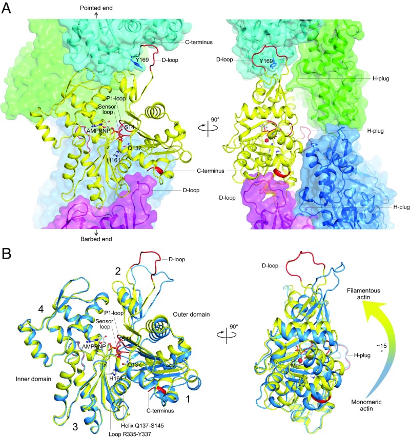
Structure of the actin filament in the AMPPNP-bound state (23). (A) Orthogonal views of the cryo-EM structure of the filament with bound AMPPNP determined at 3.1 Å resolution [Protein Data Bank (PDB) ID code 6DJM]. One subunit in the filament (yellow) interacts with four other subunits: two along the long-pitch helix (cyan, magenta) and two on the opposite strand (green, blue). The conformational change upon Pi release affects primarily the P1-loop (G13-G15), sensor loop (E72-G74), D-loop (Q41-Q49), and the C terminus (R372-F375), all highlighted red. Interactions along the long-pitch helix are stronger and involve the D-loop, which binds in the hydrophobic- (or target-)binding cleft of the subunit immediately above it, making contacts with both subdomain 1 (C terminus) and subdomain 3 (Y169). Lateral contacts involve primarily the hydrophobic plug (H-plug), which contacts two subunits on the opposite strand, including the D-loop of one of these subunits. (B) Superimposition of a subunit from the AMPPNP-bound filament structure (yellow) with the ATP-bound structure of monomeric actin in complex with a tropomodulin fragment (blue, PDB ID code 4PKG), using the inner domain as reference for fitting (G146-E334). The orientation and highlighted regions (red) are the same as in A. Numbers 1 to 4 indicate the four actin subdomains. The transition from monomeric to filamentous actin involves an ∼15° rotation of the outer domain with respect to the inner domain (arrow on the right), with helix Q137-S145 and loop R335-Y337 acting as a hinge for this rotation. This rotation repositions the side chains of Q137, H161, and the P1-loop for ATP hydrolysis.
Comment on
-
Mechanism of actin polymerization revealed by cryo-EM structures of actin filaments with three different bound nucleotides.Proc Natl Acad Sci U S A. 2019 Mar 5;116(10):4265-4274. doi: 10.1073/pnas.1807028115. Epub 2019 Feb 13. Proc Natl Acad Sci U S A. 2019. PMID: 30760599 Free PMC article.
Similar articles
-
Near-atomic resolution for one state of F-actin.Structure. 2015 Jan 6;23(1):173-182. doi: 10.1016/j.str.2014.11.006. Epub 2014 Dec 18. Structure. 2015. PMID: 25533486 Free PMC article.
-
Structural basis of actin filament capping at the barbed-end: a cryo-electron microscopy study.EMBO J. 2006 Nov 29;25(23):5626-33. doi: 10.1038/sj.emboj.7601395. Epub 2006 Nov 16. EMBO J. 2006. PMID: 17110933 Free PMC article.
-
Actin filament polymerization regulates gliding motility by apicomplexan parasites.Mol Biol Cell. 2003 Feb;14(2):396-406. doi: 10.1091/mbc.e02-08-0458. Mol Biol Cell. 2003. PMID: 12589042 Free PMC article.
-
Actin polymerization: regulation by divalent metal ion and nucleotide binding, ATP hydrolysis and binding of myosin.Adv Exp Med Biol. 1994;358:71-81. doi: 10.1007/978-1-4615-2578-3_7. Adv Exp Med Biol. 1994. PMID: 7801813 Review.
-
Mechanostress resistance involving formin homology proteins: G- and F-actin homeostasis-driven filament nucleation and helical polymerization-mediated actin polymer stabilization.Biochem Biophys Res Commun. 2018 Nov 25;506(2):323-329. doi: 10.1016/j.bbrc.2018.09.189. Epub 2018 Oct 9. Biochem Biophys Res Commun. 2018. PMID: 30309655 Review.
Cited by
-
Cryo-EM structures of cardiac muscle α-actin mutants M305L and A331P give insights into the structural mechanisms of hypertrophic cardiomyopathy.Eur J Cell Biol. 2024 Dec;103(4):151460. doi: 10.1016/j.ejcb.2024.151460. Epub 2024 Oct 1. Eur J Cell Biol. 2024. PMID: 39393252 Free PMC article.
-
Transition State of Arp2/3 Complex Activation by Actin-Bound Dimeric Nucleation-Promoting Factor.Proc Natl Acad Sci U S A. 2023 Aug 15;120(33):e2306165120. doi: 10.1073/pnas.2306165120. Epub 2023 Aug 7. Proc Natl Acad Sci U S A. 2023. PMID: 37549294 Free PMC article.
-
Conformation of actin subunits at the barbed and pointed ends of F-actin with and without capping proteins.Cytoskeleton (Hoboken). 2023 Sep-Oct;80(9-10):309-312. doi: 10.1002/cm.21770. Epub 2023 Aug 26. Cytoskeleton (Hoboken). 2023. PMID: 37632366 Free PMC article.
-
Cracked actin filaments as mechanosensitive receptors.Biophys J. 2024 Oct 1;123(19):3283-3294. doi: 10.1016/j.bpj.2024.06.014. Epub 2024 Jun 17. Biophys J. 2024. PMID: 38894540
-
Insights into Actin Isoform-Specific Interactions with Myosin via Computational Analysis.Molecules. 2024 Jun 23;29(13):2992. doi: 10.3390/molecules29132992. Molecules. 2024. PMID: 38998944 Free PMC article.
References
-
- Kabsch W, Mannherz HG, Suck D, Pai EF, Holmes KC. Atomic structure of the actin:DNase I complex. Nature. 1990;347:37–44. - PubMed
-
- Holmes KC, Popp D, Gebhard W, Kabsch W. Atomic model of the actin filament. Nature. 1990;347:44–49. - PubMed
-
- Korn ED, Carlier MF, Pantaloni D. Actin polymerization and ATP hydrolysis. Science. 1987;238:638–644. - PubMed
Publication types
MeSH terms
Substances
Grants and funding
LinkOut - more resources
Full Text Sources
