. 2019 Mar 5;116(10):4496-4501.
doi: 10.1073/pnas.1817537116. Epub 2019 Feb 19.

Dual AAV-mediated gene therapy restores hearing in a DFNB9 mouse model

Affiliations

Dual AAV-mediated gene therapy restores hearing in a DFNB9 mouse model

Omar Akil et al. Proc Natl Acad Sci U S A. .

Abstract

Autosomal recessive genetic forms (DFNB) account for most cases of profound congenital deafness. Adeno-associated virus (AAV)-based gene therapy is a promising therapeutic option, but is limited by a potentially short therapeutic window and the constrained packaging capacity of the vector. We focus here on the otoferlin gene underlying DFNB9, one of the most frequent genetic forms of congenital deafness. We adopted a dual AAV approach using two different recombinant vectors, one containing the 5' and the other the 3' portions of otoferlin cDNA, which exceed the packaging capacity of the AAV when combined. A single delivery of the vector pair into the mature cochlea of Otof-/- mutant mice reconstituted the otoferlin cDNA coding sequence through recombination of the 5' and 3' cDNAs, leading to the durable restoration of otoferlin expression in transduced cells and a reversal of the deafness phenotype, raising hopes for future gene therapy trials in DFNB9 patients.

Keywords: DFNB9; deafness; dual AAV; gene therapy; otoferlin.


Conflict of interest statement

Conflict of interest statement: W.W.H. and the University of Florida (UF) have a financial interest in the use of AAV therapies, and own equity in a company, Applied Genetic Technologies Corp. (AGTC), that might, in the future, commercialize some aspects of this work, and a joint international patent application (International Patent Application No.: PCT/US2018/031009 - WGS “Whole Genome Sequencing” Ref. U1197.70110WO00) has been submitted by UF and University of California, San Francisco (F.D., W.W.H., O.A.).

Figures

Fig. 1.
Fig. 1.
Expression of otoferlin in HEK293 cells following dual AAV-vector delivery. (Left) Schematic representation of the recombinant AAV-vector pair used in this study, and of the recombination, transcription, splicing, and translation processes producing the full-length protein otoferlin in coinfected cells. The recombinant AAV-Otof NT and AAV-Otof CT vectors contain the 5′ and 3′ parts of the otoferlin cDNA, respectively. The recombinogenic bridging sequence present in the two recombinant vectors is indicated by a gray sphere. The red bars under the protein diagram denote the two peptides used to produce the antibodies against the N-terminal and C-terminal parts of otoferlin. Abbreviations: C2, C2 domain; ITR, inverted terminal repeats; polyA, polyadenylation signal; SA, splice acceptor site; SD, splice donor site; smCBA, cytomegalovirus immediate early/chicken β-actin chimeric promoter; TM, transmembrane domain. (Right) HEK293 cells were infected with AAV-Otof NT alone (Upper), AAV-Otof CT alone (Middle), or AAV-Otof NT and AAV-Otof CT together (Lower). They were stained for otoferlin (green) with a polyclonal antibody directed against the C-terminal part of the protein 48 h later, and cell nuclei were labeled with DAPI (blue). Only coinfected cells produce otoferlin. (Scale bars, 15 μm.)
Fig. 2.
Fig. 2.
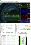
Dual AAV-mediated gene therapy in P10 Otof−/− mice restores otoferlin expression and prevents deafness. (A, Left) Confocal image of the mid-to-apical turn of the injected cochlea from a P70 mouse, immunostained for otoferlin (green). Cell nuclei are stained with DAPI (blue). A large proportion of the IHCs, but none of the outer hair cells (OHCs), express otoferlin. Arrowheads indicate nontransduced IHCs. (Inset) Higher magnification of the boxed area. (Scale bars, 50 μm and 10 μm.) (Inset). (A, Right) Images of IHCs coimmunostained for otoferlin (green), the ribbon protein ribeye (blue), and the GluA2 subunit of postsynaptic glutamate receptors (red). Synaptic active zones have a normal distribution in transduced IHCs expressing otoferlin, whereas they tend to form clusters (arrowheads) in nontransduced IHCs (indicated by dashed lines). (Scale bar, 5 μm.) (B, Left) Four weeks after the dual AAV injection, Otof−/− mice displayed ABR thresholds in response to clicks or tone bursts at frequencies of 8 kHz, 16 kHz, and 32 kHz (green dots, n = 8) close to those of wild-type mice (black dots, n = 8). By contrast, Otof−/− mice receiving AAV-Otof NT (orange dots, n = 3) or no injection (blue dots, n = 6) had no identifiable ABR waves up to sound intensity levels of 86 dB SPL. (B, Right) In the Otof−/− mice treated on P10 (arrow), the hearing thresholds for click stimuli were stable for at least 6 mo after recovery. (C, Left) ABR traces, recorded 3 wk after therapeutic injection, in a wild-type mouse (black), an Otof−/− mouse (Otof−/−, blue), and a rescued Otof−/− mouse (Otof−/− injected, green), showing similar waveforms in the wild-type and rescued mice. (C, Right) Bar graph showing the latency and normalized amplitude of ABR wave I for 86 dB SPL click stimuli in rescued Otof−/− mice (green, n = 8) and wild-type mice (black, n = 5). Average maximum value of wave I amplitude, measured at 86 dB SPL, was 4.6 ± 0.9 μV and 1.8 ± 0.1 μV in Otof +/+ mice and treated Otof−/− mice, respectively.
Fig. 3.
Fig. 3.
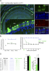
Dual AAV-mediated gene therapy in Otof−/− mice on P17 durably restores otoferlin expression and hearing. (A, Left) Confocal image of the mid-to-apical turn of the injected cochlea from a P80 mouse, immunostained for otoferlin (green). Cell nuclei are stained with DAPI (blue). Most IHCs express otoferlin, whereas outer hair cells (OHCs) do not. Arrowheads indicate nontransduced IHCs. (Inset) Higher magnification of the boxed area. (Scale bars, 50 μm and 10 μm.) (Inset). (A, Right) Images of IHCs coimmunostained for otoferlin (green), the ribbon protein ribeye (blue), and the GluA2 subunit of postsynaptic glutamate receptors (red). Synaptic active zones have a normal distribution in transduced IHCs expressing otoferlin, whereas they tend to form clusters (arrowheads) in nontransduced IHCs (indicated by dashed lines). (Scale bar, 5 μm.) (B, Left) ABR thresholds of untreated Otof−/− mice (blue, n = 5), treated Otof−/− mice (green, n = 5), and wild-type mice (black, n = 5) in response to clicks or tone-burst stimuli at frequencies of 8, 16, and 32 kHz, 4 wk after intracochlear injection of the recombinant vector pair in the treated mice. (B, Right) Time course of hearing recovery in Otof−/− mice receiving injections on P17 (arrow). Hearing restoration to near-wild-type levels is maintained for at least 20 wk postinjection. (C, Left) ABR traces, recorded 2 wk after therapeutic injection, in a wild-type mouse (black), an Otof−/− mouse (Otof−/−, blue), and a rescued Otof−/− mouse (Otof−/− injected, green), showing similar waveforms in the wild-type and rescued mice. (C, Right) Bar graph showing that the latency of ABR wave I (for 86 dB SPL click stimuli) in rescued Otof−/− mice (n = 5) is similar to that in wild-type mice (n = 5), whereas its normalized amplitude is about half that in wild-type mice (average maximum value of wave I amplitude, measured at 86 dB SPL, was 4.6 ± 0.9 μV and 2.2 ± 0.4 μV in Otof +/+ mice and treated Otof−/− mice, respectively).
Fig. 4.
Fig. 4.
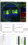
Dual AAV-mediated gene therapy in Otof−/− mice on P30 restores otoferlin expression and hearing in a sustained manner. (A, Left) Confocal image of the mid-to-apical turn of the injected cochlea from a P40 mouse, immunostained for otoferlin (green). Cell nuclei are stained with DAPI (blue). Most IHCs express otoferlin, whereas outer hair cells (OHCs) do not. Arrowheads indicate nontransduced IHCs. (Inset) Higher magnification of the boxed area. (Scale bars, 50 μm and 10 μm.) (Inset). (A, Right) Images of IHCs coimmunostained for otoferlin (green), the ribbon protein ribeye (blue), and the GluA2 subunit of postsynaptic glutamate receptors (red). Synaptic active zones have a normal distribution in transduced IHCs expressing otoferlin, whereas they tend to form clusters (arrowheads) in nontransduced IHCs (indicated by dashed lines). (Scale bar, 5 μm.) (B) ABR thresholds of untreated Otof−/− mice (blue, n = 3), treated Otof−/− mice (green, n = 3), and wild-type mice (black, n = 3) in response to clicks or tone-burst stimuli at frequencies of 8, 16, and 32 kHz, 3 wk (Left), 14 wk, and 20 wk (Right) after intracochlear injection of the recombinant vector pair in the treated mice. In these mice hearing restoration to near-wild-type levels is maintained for at least 20 wk postinjection. (C, Left) ABR traces, recorded 7 wk after therapeutic injection, in a wild-type mouse (black), an Otof−/− mouse (Otof−/−, blue), and a rescued Otof−/− mouse (Otof−/− injected, green), showing similar waveforms in the wild-type and rescued mice. (C, Right) Bar graph showing that the latency of ABR wave I (for 86 dB SPL click stimuli) in rescued Otof−/− mice (n = 3) is similar to that in wild-type mice (n = 3), whereas its normalized amplitude is about half that in wild-type mice (average maximum value of wave I amplitude, measured at 86 dB SPL, was 4.6 ± 0.9 μV and 2.5 ± 0.1 μV in Otof +/+ mice and treated Otof−/− mice, respectively).

References

    1. Duman D, Tekin M. Autosomal recessive nonsyndromic deafness genes: A review. Front Biosci. 2012;17:2213–2236. - PMC - PubMed
    1. Kral A, O’Donoghue GM. Profound deafness in childhood. N Engl J Med. 2010;363:1438–1450. - PubMed
    1. Jeon EK, Turner CW, Karsten SA, Henry BA, Gantz BJ. Cochlear implant users’ spectral ripple resolution. J Acoust Soc Am. 2015;138:2350–2358. - PMC - PubMed
    1. Vivero RJ, Fan K, Angeli S, Balkany TJ, Liu XZ. Cochlear implantation in common forms of genetic deafness. Int J Pediatr Otorhinolaryngol. 2010;74:1107–1112. - PMC - PubMed
    1. Flotte TR, et al. Phase I trial of intramuscular injection of a recombinant adeno-associated virus alpha 1-antitrypsin (rAAV2-CB-hAAT) gene vector to AAT-deficient adults. Hum Gene Ther. 2004;15:93–128. - PubMed

Publication types