. 2019 Apr 19;294(16):6214-6226.
doi: 10.1074/jbc.RA118.006876. Epub 2019 Feb 19.

Thermodynamic insights into an interaction between ACYL-CoA-BINDING PROTEIN2 and LYSOPHOSPHOLIPASE2 in Arabidopsis

Affiliations

Thermodynamic insights into an interaction between ACYL-CoA-BINDING PROTEIN2 and LYSOPHOSPHOLIPASE2 in Arabidopsis

Rui Miao et al. J Biol Chem. .

Abstract

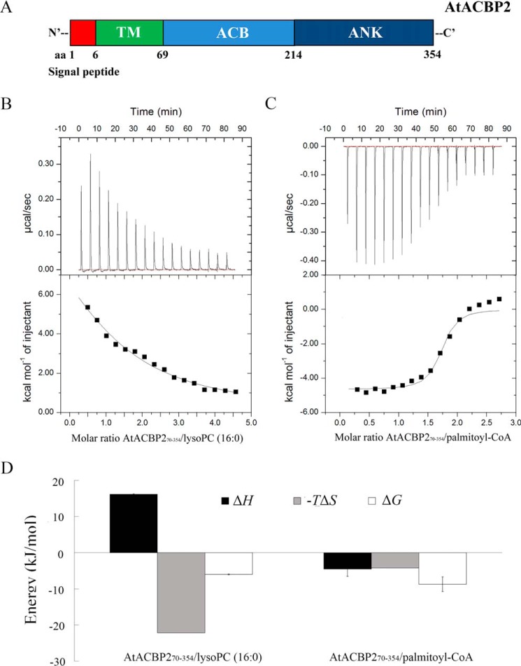
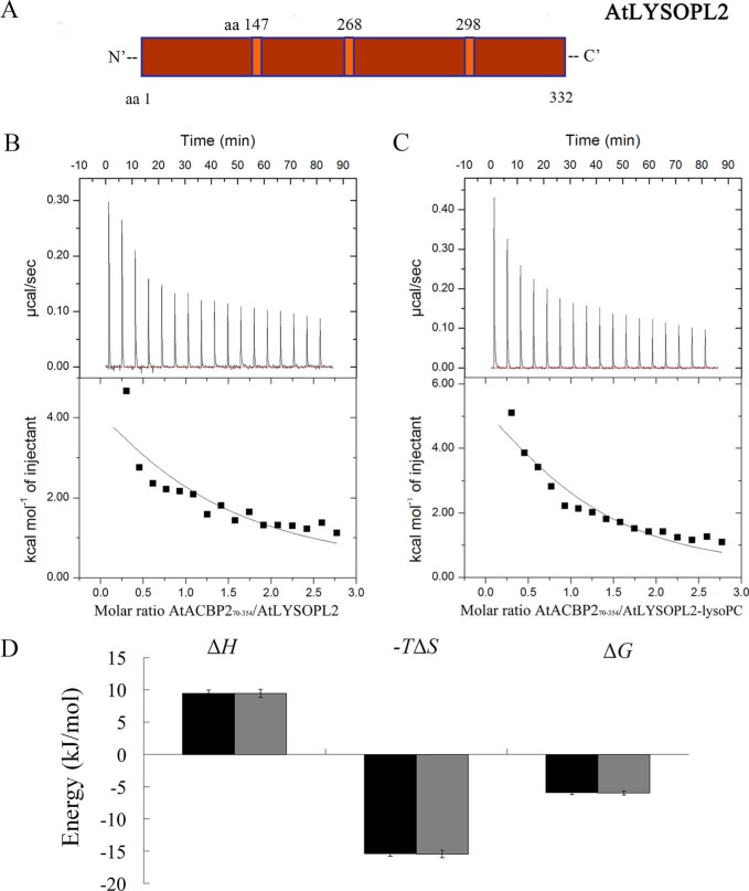
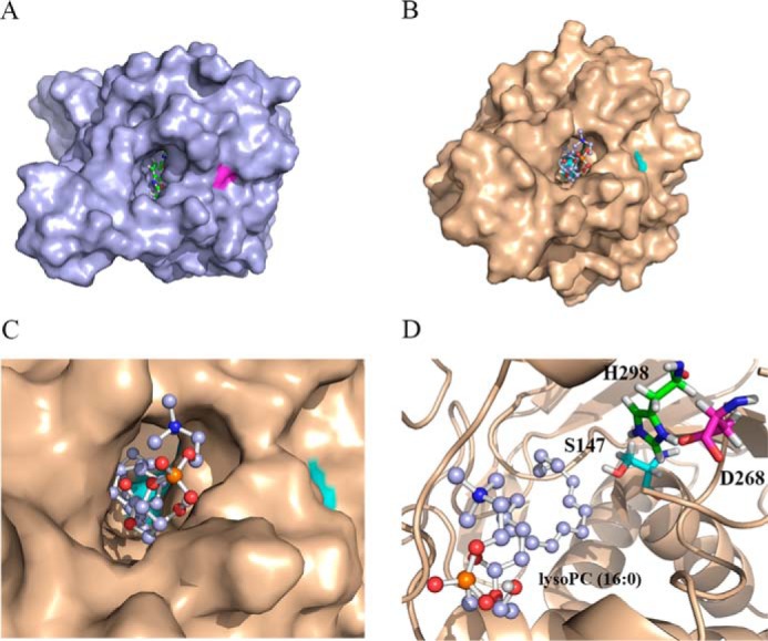
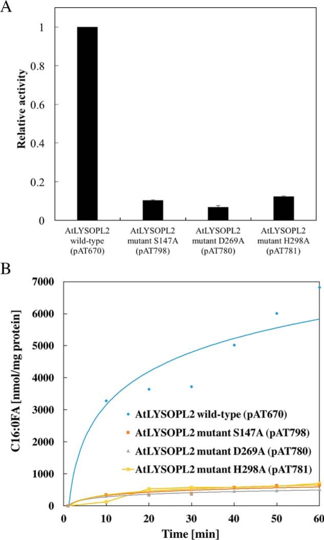
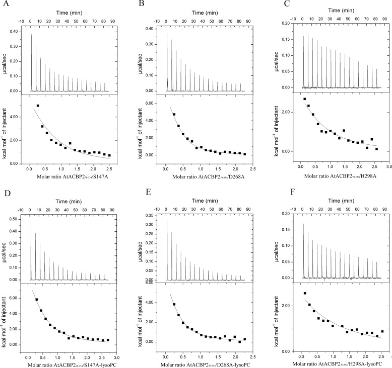
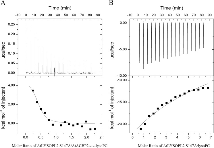
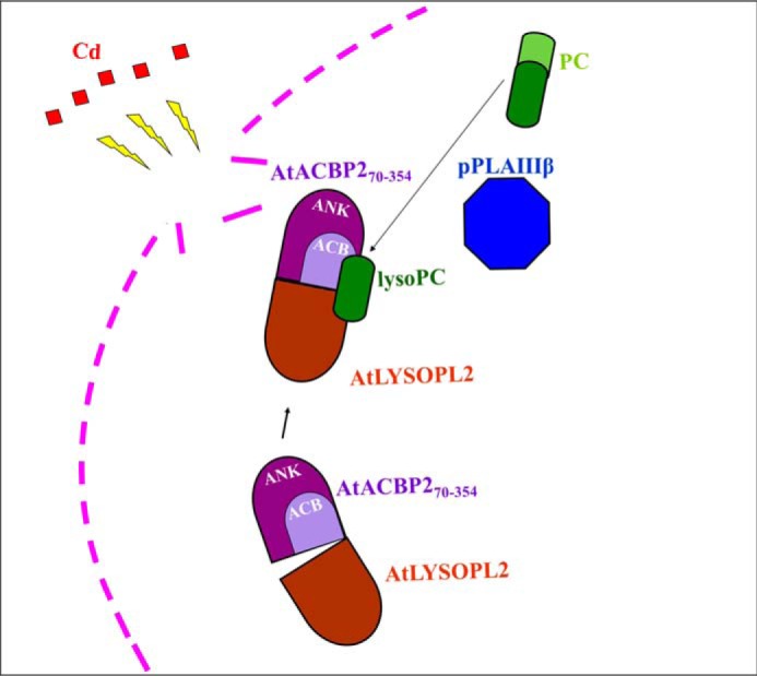
Lysophospholipids (LPLs) are important lipid-signaling molecules in plants, of which lysophosphatidylcholine (lysoPC) is one of the most well-characterized LPLs, having important roles in plant stress responses. It is broken down by lysophospholipases, but the molecular mechanism involved in lysoPC degradation is unclear. Recombinant Arabidopsis thaliana ACYL-CoA-BINDING PROTEIN2 (AtACBP2) has been reported to bind lysoPC via its acyl-CoA-binding domain and also LYSOPHOSPHOLIPASE 2 (AtLYSOPL2) via its ankyrin repeats in vitro To investigate the interactions of AtACBP2 with AtLYSOPL2 and lysoPC in more detail, we conducted isothermal titration calorimetry with AtACBP270-354, an AtACBP2 derivative consisting of amino acids 70-354, containing both the acyl-CoA-binding domain and ankyrin repeats. We observed that the interactions of AtACBP270-354 with AtLYSOPL2 and lysoPC were both endothermic, favored by solvation entropy and opposed by enthalpy, with dissociation constants in the micromolar range. Of note, three AtLYSOPL2 catalytic triad mutant proteins (S147A, D268A, and H298A) bound lysoPC only weakly, with an exothermic burst and dissociation constants in the millimolar range. Furthermore, the binding affinity of lysoPC-premixed AtACBP270-354 to AtLYSOPL2 was 10-fold higher than that of AtACBP270-354 alone to AtLYSOPL2. We conclude that AtACBP2 may play a role in facilitating a direct interaction between AtLYSOPL2 and lysoPC. Our results suggest that AtACBP270-354 probably binds to lysoPC through a hydrophobic interface that enhances a hydrotropic interaction of AtACBP270-354 with AtLYSOPL2 and thereby facilitates AtLYSOPL2's lysophospholipase function.

Keywords: ankyrin; enzyme mutation; isothermal titration calorimetry (ITC); lipid metabolism; lysophosphatidylcholine; lysophospholipid; molecular docking; protein–protein interaction; structural model; thermodynamics.


Conflict of interest statement

The authors declare that they have no conflicts of interest with the contents of this article

Figures

Figure 1.
Figure 1.
ITC analysis of AtACBP270–354 interactions with lysoPC (C16:0) and C16:0–CoA. A, schematic representation of the domains in Arabidopsis AtACBP2. The signal peptide (aa 1–6), transmembrane (TM) domain (aa 7–69), acyl-CoA binding (ACB) domain (aa 70 to 214), and ankyrin repeats (ANK) (aa 215–354) of AtACBP2 are shown in red, green, light blue, and dark blue, respectively. AtACBP270–354 consists of a derivative lacking the transmembrane domain, with the ACB and ANK domains intact. AtACBP2215–354 consists of the ANK domain. B, AtACBP270–354 and lysoPC (C16:0) binding measured by titrating 30–40 μm AtACBP270–354 in the chamber with 600–800 μm lysoPC (C16:0) in the syringe. C, AtACBP270–354 and C16:0–CoA binding measured by titrating 30–40 μm AtACBP270–354 in the chamber with 600–800 μm C16:0–CoA in the syringe. Top panel, raw heating power over time; bottom panel, fit of the integrated energy values normalized for injected protein. D, binding signature (ΔH, −TΔS, and ΔG) plotted for AtACBP270–354–lysoPC (C16:0) and AtACBP270–354–C16:0–CoA interactions. Binding enthalpy, entropy, and free energy are shown in black, gray, and white, respectively. The values originate from Table 1. Error bars denote S.E., n = 2.
Figure 2.
Figure 2.
ITC analysis of AtLYSOPL2, AtACBP270–354, and lysoPC interactions. A, schematic representation of the structure of Arabidopsis AtLYSOPL2. The catalytic triad of AtLYSOPL2 is shown in orange. B, interactions between AtLYSOPL2 and AtACBP270–354. Binding was tested in ITC by titrating 30–40 μm AtLYSOPL2 in the chamber with 400–500 μm AtACBP270–354 in the syringe. C, interactions between the AtLYSOPL2–lysoPC (molar ratio 1:1) complex and AtACBP270–354. Binding was tested in ITC by titrating 30 μm AtLYSOPL2–lysoPC complex in the chamber with 400–500 μm AtACBP270–354 in the syringe. Top panel, raw heating power over time; bottom panel, fit of the integrated energy values normalized for injected protein. D, energetics of AtLYSOPL2 interaction with AtACBP270–354. Plots of the binding signature (ΔH, −TΔS, and ΔG) with interactions of AtLYSOPL2 and AtACBP270–354 in the absence of lysoPC shown in black, and AtLYSOPL2 and AtACBP270–354 in the presence of lysoPC in gray. The values were obtained from data presented in Table 2. Error bars denote S.E., n = 3. The values originate from Table 2.
Figure 3.
Figure 3.
Homology modeling and dock simulation of lysoPC (C16:0) binding to AtLYSOPL2. A, structure of human MGL with its inhibitor JZL184 (Protein Data Bank code 3JWE (21). B and C, 3D structure model of AtLYSOPL2 with lysoPC (C16:0). The bottom catalytic residues in the pocket-like cleavage site are shown in cyan. D, putative scissor aa residues Ser-147, Asp-268, and His-298 in the cleavage site of AtLYSOPL2. LysoPC (C16:0) is shown as ball and stick.
Figure 4.
Figure 4.
Lysophospholipase activity assays on AtLYSOPL2 and its mutant derivatives. A, comparison of lysophospholipase activity among WT AtLYSOPL2 and its mutant proteins (S147A, D268A, and H298A). The values are shown as the relative activity to WT AtLYSOPL2 (defined value of 1). B, amount of C16:0-FA detected at different time intervals.
Figure 5.
Figure 5.
ITC analysis of interactions between AtACBP270–354 and AtLYSOPL2 mutant proteins in the absence or presence of lysoPC. A–C, AtACBP270–354interactions with AtLYSOPL2 mutants S147A (A), D268A (B), and H298A (C) in the absence of lysoPC. Interactions were measured by titrating 20–30 μm AtLYSOPL2 (S147A, D268A, and H298A) in the chamber with 300–400 μm AtACBP270–354 in the syringe. D–F, AtACBP270–354 interactions with AtLYSOPL2 mutants S147A (D), D268A (E), H298A (F) in the presence of lysoPC. Interactions were measured by titrating 30 μm AtLYSOPL2 (S147A, D268A, and H298A)–lysoPC complex (molar ratio 1:1) in the chamber with 400–500 μm AtACBP270–354 in the syringe. Top panel, raw heating power over time; bottom panel, fit of the integrated energy values normalized for the injected protein. The values originate from Table 2.
Figure 6.
Figure 6.
ITC analysis of AtLYSOPL2 catalytic triad mutant S147A interactions with the AtACBP270–354–lysoPC complex and lysoPC. A, binding of AtLYSOPL2 S147A and AtACBP270–354–lysoPC complex was measured by titrating 20–30 μm AtLYSOPL2 S147A in the chamber with 400–500 μm of the AtACBP270–354–lysoPC (molar ratio 1:1) complex in the syringe. B, AtLYSOPL2 S147A and lysoPC interaction was measured by titrating 20–30 μm AtLYSOPL2 S147A in the chamber with 600–800 μm lysoPC in the syringe. Top panel, raw heating power over time; bottom panel, fit of the integrated energy values normalized for the injected protein. The values originate from Tables 3 and 4.
Figure 7.
Figure 7.
Proposed working model for AtLYSOPL2, AtACBP2, and lysoPC interaction. LysoPC, generated by pPLAIIIβ action on phosphatidylcholine (PC), binds to AtACBP2 and promotes the formation of an AtLYSOPL2–AtACBP2 complex. This complex could improve efficiency in membrane repair following Cd-induced stress. Cd, red; plasma membrane, pink; lysoPC, dark green; PC, light green; ankyrin-repeat (ANK) domain, purple; acyl-CoA-binding (ACB) domain, light purple; PLAIII β, blue; AtLYSOPL2, brown.

References

    1. de Kroon A. I., Rijken P. J., and De Smet C. H. (2013) Checks and balances in membrane phospholipid class and acyl chain homeostasis, the yeast perspective. Prog. Lipid Res. 52, 374–394 10.1016/j.plipres.2013.04.006 - DOI - PubMed
    1. Grechkin A. (1998) Recent developments in biochemistry of the plant lipoxygenase pathway. Prog. Lipid Res. 37, 317–352 10.1016/S0163-7827(98)00014-9 - DOI - PubMed
    1. Wang X. (2004) Lipid signaling. Curr. Opin. Plant Biol. 7, 329–336 10.1016/j.pbi.2004.03.012 - DOI - PubMed
    1. Ryu S. B. (2004) Phospholipid-derived signaling mediated by phospholipase A in plants. Trends Plant Sci. 9, 229–235 10.1016/j.tplants.2004.03.004 - DOI - PubMed
    1. Welti R., Li W., Li M., Sang Y., Biesiada H., Zhou H. E., Rajashekar C. B., Williams T. D., and Wang X. (2002) Profiling membrane lipids in plant stress responses: role of phospholipase Dα in freezing-induced lipid changes in Arabidopsis. J. Biol. Chem. 277, 31994–32002 10.1074/jbc.M205375200 - DOI - PubMed

Publication types

MeSH terms

LinkOut - more resources